
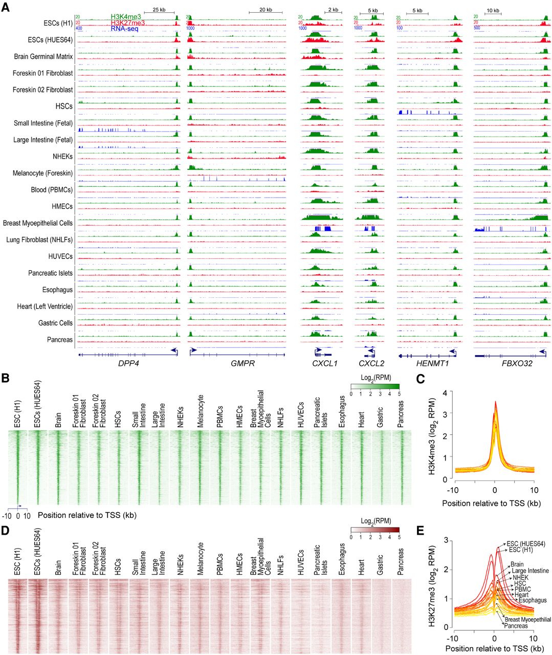
H3K4me3, observed at bivalent promoters in ESCs, persists in nearly all cell types irrespective of gene expression. (A) Genome browser shots of select genes, bivalently marked in human embryonic stem cells (ESCs), showing ChIP-seq read density profiles for H3K4me3 (green) and H3K27me3 (red) in various cell types. Also shown are read density profiles for gene expression (blue; RNA-seq). (HSCs) hematopoietic stem cells; (PBMCs) peripheral blood mononuclear cells; (HMECs) human mammary epithelial cells; (NHEKs) normal human embryonic kidneys; (NHLFs) normal human lung fibroblasts; (HUVECs) human umbilical vein endothelial cells. (B,D) Heatmap representation of H3K4me3 (B) and H3K27me3 (D) ChIP-seq read density, in various cell types, near transcription start sites (TSSs) of genes bivalently marked in human ESCs. Genes were ordered by decreasing order of H3K4me3 signal in ESCs (top to bottom). Read density is represented as reads per million mapped reads (RPKM). (C,E) Average H3K4me3 (C) and H3K27me3 (E) ChIP-seq read density, in various cell types, near TSSs of genes shown in B. Shades of color represent individual cell types. Select cell types, of the 20 plotted, are highlighted.


