
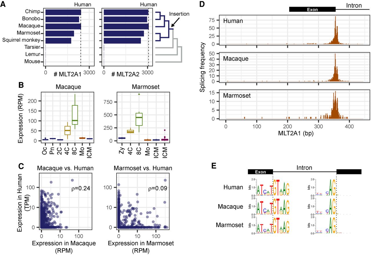
Activation of MLT2A1 and MLT2A2 elements in primate embryos. (A) Numbers of MLT2A1 and MLT2A2 elements in seven primate genomes and the mouse genome. Numbers of human MLT2A elements are denoted by dashed lines. The phylogenetic tree is shown on the right side. (B) Box plots of normalized expression values (reads per million mapped reads [RPM]) of MLT2A1 elements: (Oc) oocyte; (Pn) pronuclei; (2C, 4C, and 8C) two-cell, four-cell, and eight-cell embryos, respectively; (Mo) morula; (ICM) inner cell mass. (C) Normalized expression values of individual MLT2A1 elements in macaque versus human and marmoset versus human embryos. The numbers of elements robustly transcribed in two species (more than 5 TPM and 5 RPM) are seven for macaque versus human and 13 for marmoset versus human: (ρ) Spearman's correlation coefficients. (D) Frequency of splicing events in MLT2A1 elements of human, macaque, and marmoset. (E) Sequence logos of splice sites in MLT2A1 elements found in four-cell and eight-cell embryos of human, macaque, and marmoset. The left side shows donor sites inside MLT2A1, and the right side shows acceptor sites outside MLT2A1.


