
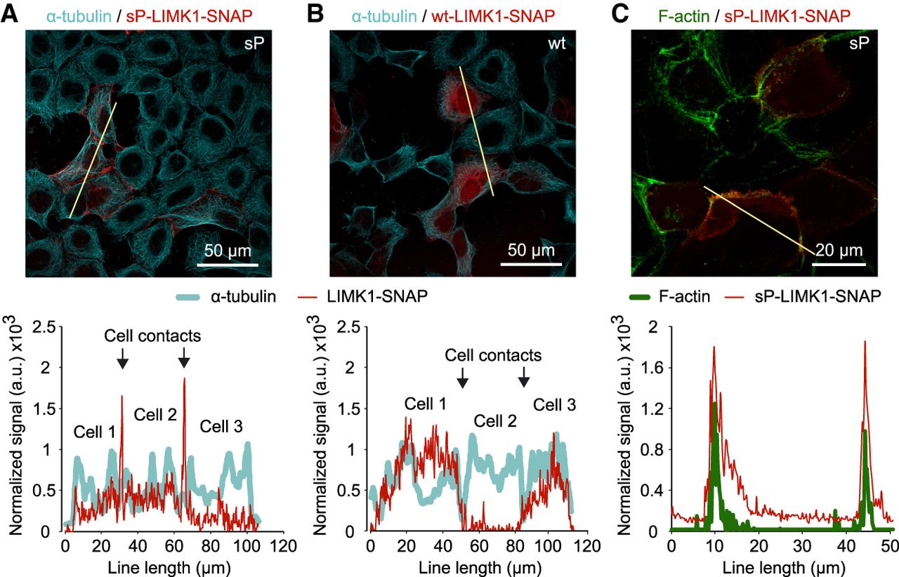
Figure 5.
Localization of LIMK1-SNAP-derived products in cells. (A,B) Products from sP and wt constructs, respectively (red, stained with SNAP-Cell 647-SiR), counterstained with α-tubulin/Alexa Fluor 488 antibodies (cyan). (Bottom) Line profile analysis across three contacting cells shows the predominantly peripheral distribution of sP products, in contrast to more uniform cytosolic distribution of the wt products. (C) Colocalization of sP products (red) and F-actin (green, stained with phalloidin-Alexa 546). Images represent stacks of four (A,B) or three (C) focal planes taken with a 5 µm step.


