
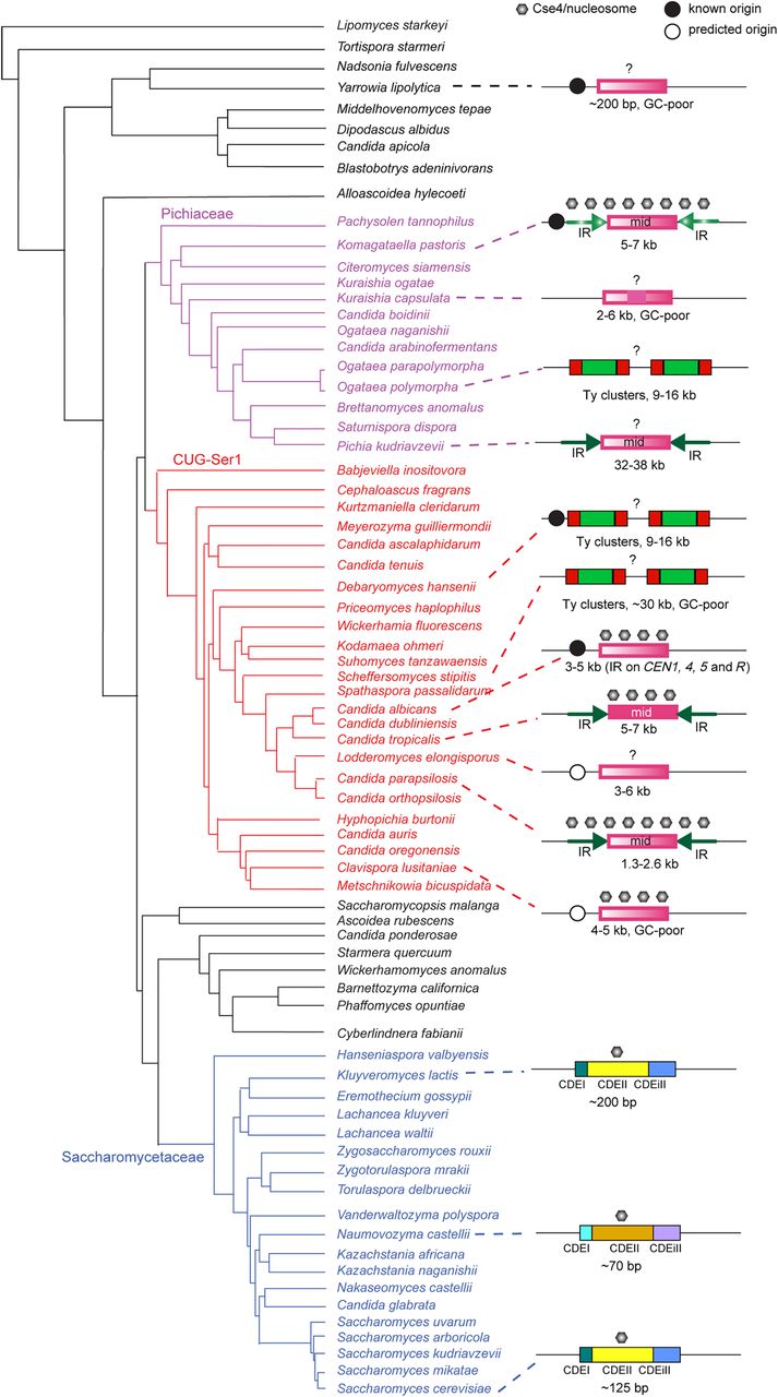
Organization of centromeres in Saccharomycotina species. The phylogeny is adapted from Shen et al. (2016). The size indicated on the centromeres refers to the region bound by Cse4 when known, or else when predicted bioinformatically, except for the Saccharomycetaceae, for which the size of the point centromere is shown. Solid color indicates conservation of sequence across centromeres in the same species, whereas a color gradient indicates unique sequences. IRs are shown with arrows; Ty clusters, as red and green boxes. Black circles show known (solid) or predicted (open) early-firing origins of replication (for details, see text). Point centromeres are conserved across the Saccharomycetaceae except for the Naumovozyma lineage, which has different sequences. Question marks indicate that localization of Cse4 nucleosomes has not been determined.


