
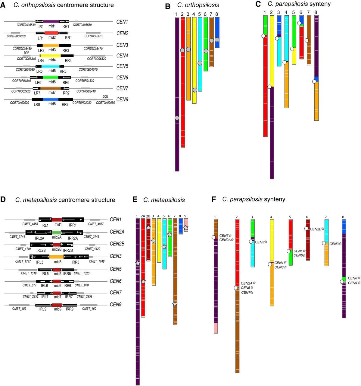
Identification of centromeres and centromere-proximal rearrangements in C. orthopsilosis and C. metapsilosis. (A) Cartoon of centromere structure in C. orthopsilosis 90-125 (Lombardi et al. 2019b). All mid regions are unique and are shown in different colors. Sequences in black are conserved among chromosomes. IRs are shown with white arrows, and adjacent genes are shown with gray boxes. Putative transposases with DDE domains are indicated. More detail is provided in Supplemental Figure S2 and Supplemental Table S2. (B,C) Synteny relationship between C. parapsilosis and C. orthopsilosis. SynChro (Drillon et al. 2014) was used (delta value of two) to identify potential orthologs (reciprocal best hits [RBHs]), represented by colored lines in the two species, and to generate synteny maps. (B) Location of RBHs on C. orthopsilosis chromosomes. The approximate location of the putative centromeres is indicated with a gray polygon. (C) C. parapsilosis chromosomes, colored with respect to the RBH from C. orthopsilosis. The location of the C. parapsilosis centromeres are indicated with an offset white circle. The location of syntenic C. orthopsilosis centromeres is shown in more detail in Figure 5. (D) Cartoon of centromere structure in C. metapsilosis. Sequences in black are conserved among chromosomes. IRs are shown with white arrows, which are sometimes fragmented and overlapping. Mid-core regions from some CENs are similar in sequence (>60%) and are shown in the same color. Adjacent genes are shown with gray boxes. More detail is provided in Supplemental Figure S2 and Supplemental Table S2. (E,F) Synteny relationship between C. parapsilosis and C. metapsilosis. (E) Location of RBHs on C. metapsilosis chromosomes. The approximate location of the putative C. metapsilosis centromeres are indicated with a gray star (centromeres were not identified on scaffolds 4 and 8). (F) C. parapsilosis chromosomes, colored with respect to the RBH from C. metapsilosis. The location of the C. parapsilosis centromeres are indicated with a white circle. The approximate location of syntenic C. metapsilosis centromeres are shown by name and with gray stars. The same colors are used for C. orthopsilosis (B) and C. metapsilosis (E). This does not indicate that synteny is completely conserved between these species; it is a feature of SynChro, which carries out pairwise comparisons.


