
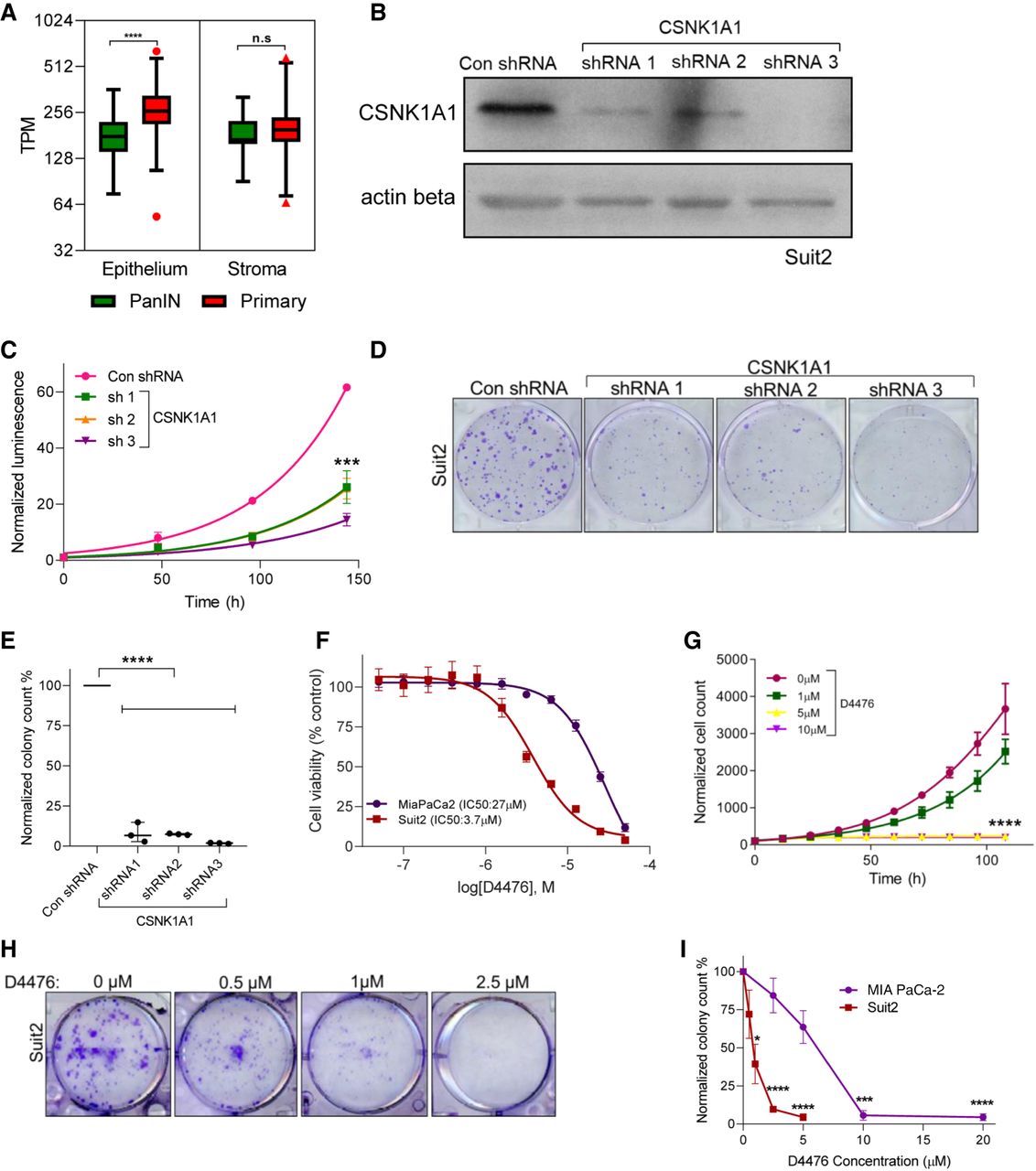
CSNK1A1 is required for cell proliferation and is a putative drug target in PDAC. (A) A plot showing CSNK1A1 gene expression (in transcripts per million) in PDAC (red) as compared to PanIN lesions (green) in the epithelium and stroma from microdissected samples: (****) P < 0.001. (B) A representative blot confirming CSNK1A1 knockdown in Suit2 cells with a nontargeting control shRNA (Con shRNA) or with one of three different shRNAs targeting CSNK1A1: n = 3. ACTB (also known as actin beta) is shown as a loading control. (C) Cell proliferation of Suit2 control and CSNK1A1-knockdown cells: n = 3; (***) P < 0.005. (D) Clonogenic growth assay of control and CSNK1A1-knockdown cells: n = 3. (E) Quantification shows the number of colonies in D: n = 3; (****) P < 0.001. (F) Dose-response of MIA PaCa-2 (purple) and Suit2 (red) cell lines to the CSNK1A1 small molecule inhibitor, D4476: n = 3. (G) Cell proliferation of Suit2 cells treated with indicated doses of D4476: n = 3; (****) P < 0.001. (H) Clonogenic growth assay of Suit2 cells treated with indicated drug doses. (I) Quantification shows the number of colonies in H: (***) P < 0.005; (****) P < 0.001.


