
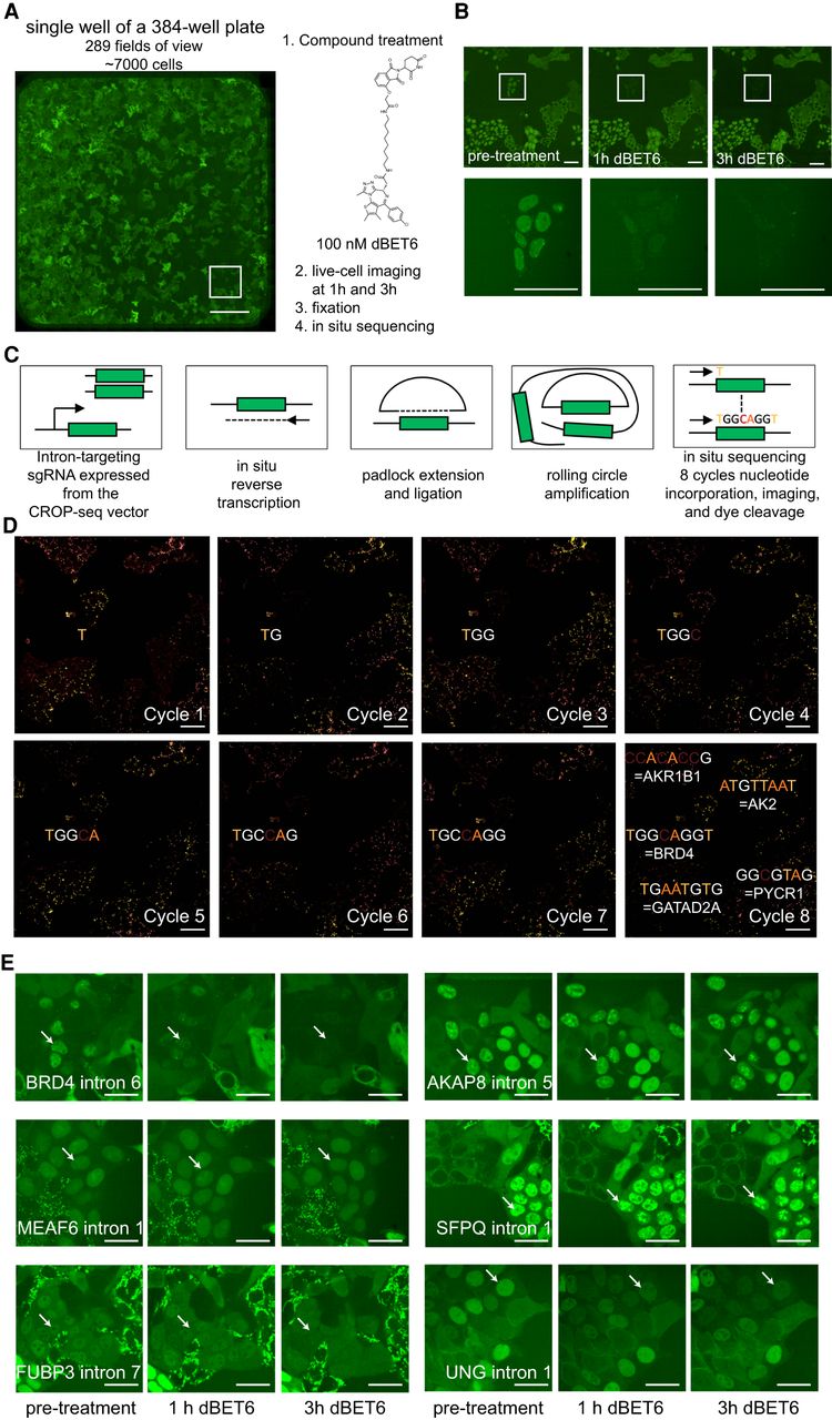
Compound screening on cell pools followed by in situ sequencing enables the detection of protein-specific compound effects. (A) Stitched image of 289 fields of view representing an entire well on a 384-well plate containing approximately 7000 individual cells. Scale bar, 500 µm. (B) Identification of a clone with rapid loss of GFP signal following treatment with 100 nM dBET6, whereas neighboring clones are unaffected. Scale bars, 50 µm. (C) Outline of the in situ sequencing approach. (D) Images from eight cycles in situ sequencing of the area shown in panel B. Scale bar, 25 µm. (E) Selected images for cells within the pool showing localization changes following dBET6 treatment. Scale bars, 25 µm.


