
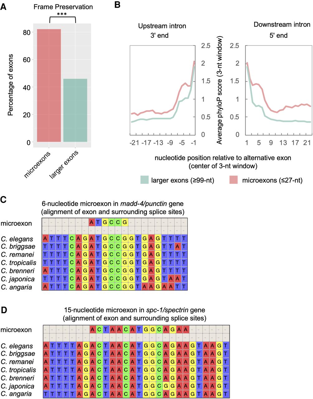
Figure 9.
Microexons <27 nt are highly conserved in Caenorhabditis species. (A) Comparison of frame preservation frequency among microexons (pink) and larger exons (green). (***) P < 1 × 10⁻¹⁰, Pearson's chi-squared test. (B) Plot of average phyloP score (rolling 3-nt average) over last 23 nt (left panel) and first 23 nt (right panel) of the upstream and downstream introns flanking microexons (pink) or larger exons (green), respectively. (C) A multiple sequence alignment displaying a highly conserved 6-nt microexon and its surrounding splice sites in the madd-4 gene. (D) A multiple sequence alignment of a highly conserved 15-nt microexon and its surrounding splice sites in the spc-1 gene.


