
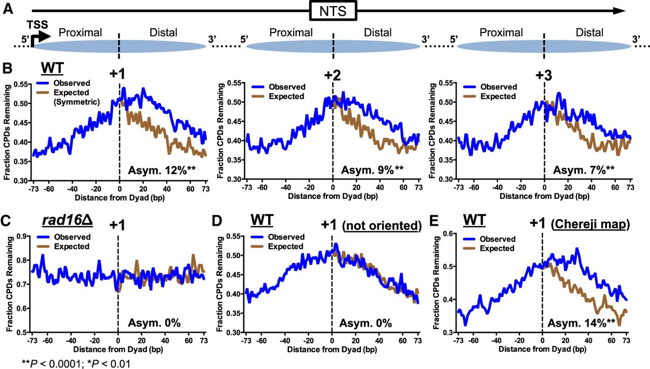
Repair of the NTS is asymmetric in yeast nucleosomes. (A) Diagram depicting how the NTS associated with each intragenic nucleosome (i.e., +1, +2, and +3) was oriented in the 5′-to-3′ direction, so that the 5′ side (i.e., TSS-proximal side) is consistently to the left of the dyad axis, whereas the 3′ side (i.e., TSS-distal side) is to the right of the dyad axis. Arrow indicates the TSS and direction of transcription. Dashed lines indicate the dyad axis of the +1, +2, and +3 nucleosomes. (B) Repair of CPD lesions in the +1 (left), +2 (middle), and +3 nucleosomes (right) in yeast genes. The fraction of CPDs remaining at each position in the nucleosome (−73 to +73 bp relative to the dyad axis) is plotted for the WT 2-h repair sample, relative to WT 0-h sample. “Observed” plots the actual CPD repair data, whereas “Expected” plots the data from the 5′ side of the nucleosome on the 3′ side (i.e., the expected fraction of CPDs remaining if repair was symmetric across the nucleosome dyad). The relative difference between the observed and expected curves quantifies the degree of asymmetry (Asym.) in repair, which is expressed as percent value (Methods). Nucleosome dyad positions were obtained from Weiner et al. (2015). (C) Same as B, left panel (+1 nucleosome), except the CPD-seq data for the rad16Δ strain were analyzed. (D) Same as B, except the NTS was not oriented in a 5′-to-3′ direction. (E) Same as B, except the +1 nucleosome dyad positions were obtained from Chereji et al. (2018).


