
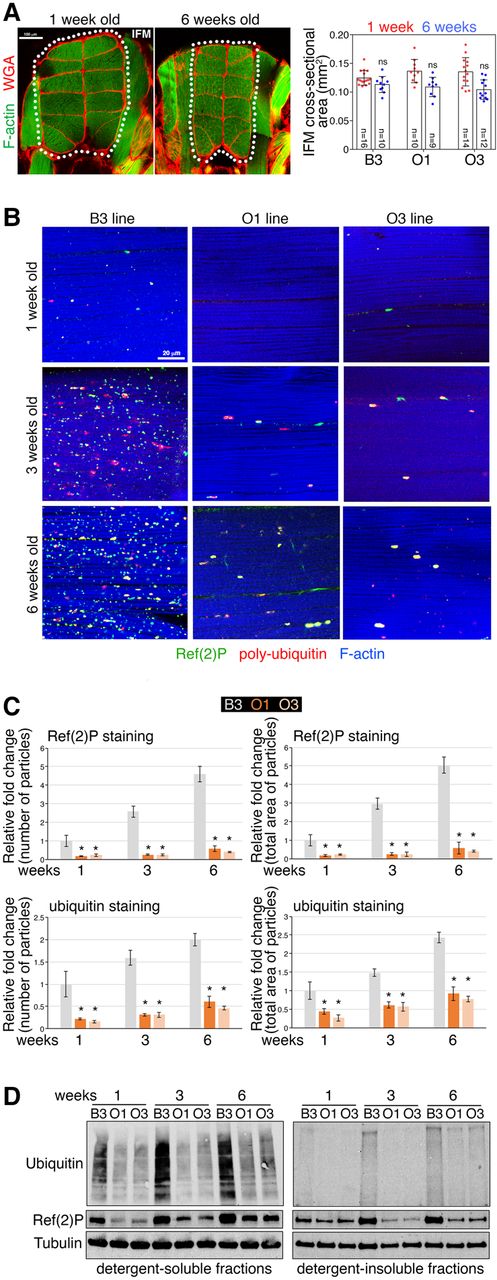
Modulation of muscle cross-sectional area and protein quality control in O and B lines during aging. (A) Changes in muscle size are variable and typically not significant during Drosophila aging. Sections of Drosophila thoraces from B3 flies at 1 and 6 wk stained with phalloidin, to detect F-actin (green), and with Alexa555-WGA (wheat germ agglutinin), to detect the extracellular matrix (red). Age-related changes in the cross-sectional area of the 12 longitudinal indirect flight muscles (IFM; outlined) are not statistically significant due to high variability (SD, n = 9–16). These findings indicate that muscle mass loss is not a robust feature of aging in Drosophila and that it does not differ between O and B lines. (B) Immunostaining of indirect flight muscle from the O and B lines reveals an age-related accumulation of Ref(2)P-positive (green) poly-ubiquitin (red) protein aggregates in the muscles of B3 flies, whereas this does not occur to the same extent in the O lines, as demonstrated by the quantifications shown in C, which report the number and total area of particles (protein aggregates) normalized by tissue area; (*) P < 0.05; n = 10–25; SEM. (D) Western blots of skeletal muscle fractions similarly indicate that there is a lower increase with aging of poly-ubiquitin protein aggregates in the muscles of the O lines compared to the B3 (ubiquitin and Ref[2]P signals detected in detergent-insoluble fractions), whereas there is no change in the ubiquitin and Ref(2)P levels detected in the detergent-soluble fractions.


