
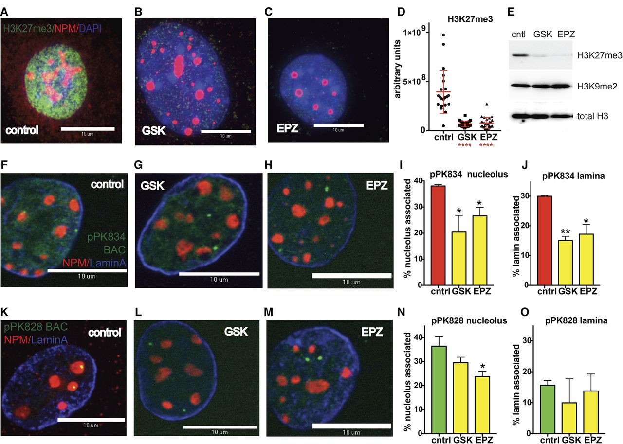
Inhibition of H3K27 methylation blocks NAD localization (A) Immunofluorescence measurement of H3K27me3 levels in control cells. (B) As in A, except cells were treated with GSK126. (C) As in A, except cells were treated with EPZ6438. (D) Quantitation of IF data from A–C. GSK126: (****) P < 0.0001, Welch's t-test; EPZ6438: (****) P < 0.0001, Welch's t-test. (E) Immunoblot analyses of cells treated as in A–C. (F) 3D immuno-FISH localization of Type I probe pPK834 (green), anti-nucleophosmin antibodies (NPM; ab10530 [Abcam], red), and Anti-Lamin A antibodies (blue). Untreated control cells. (G) As in F, except that cells were treated with GSK126. (H) As in F, except that cells were treated with EPZ6438. (I) Quantitation of pPK834 nucleolar association measured by DNA-FISH as in Figure 3. GSK126: (*) P = 0.040, Welch's t-test; EPZ6438: (*) P = 0.023, Welch's t-test. (J) Quantitation of pPK834 laminar association measured by DNA-FISH as in Figure 3. GSK126: (**) P = 0.0029, Welch's t-test; EPZ6438: (*) P = 0.021, Welch's t-test. (K) 3D immuno-FISH localization of Type II probe pPK828 (green), with other markers as in A. Untreated control cells. (L) As in K, except that cells were treated with GSK126. (M) As in K, except that cells were treated with EPZ6438. (N) Quantitation of pPK828 nucleolar association. GSK126: P = 0.083, Welch's t-test; EPZ6438: P = 0.018, Welch's t-test. (O) Quantitation of pPK828 laminar association. GSK126: P = 0.33, Welch's t-test; EPZ6438: P = 0.63, Welch's t-test.


