
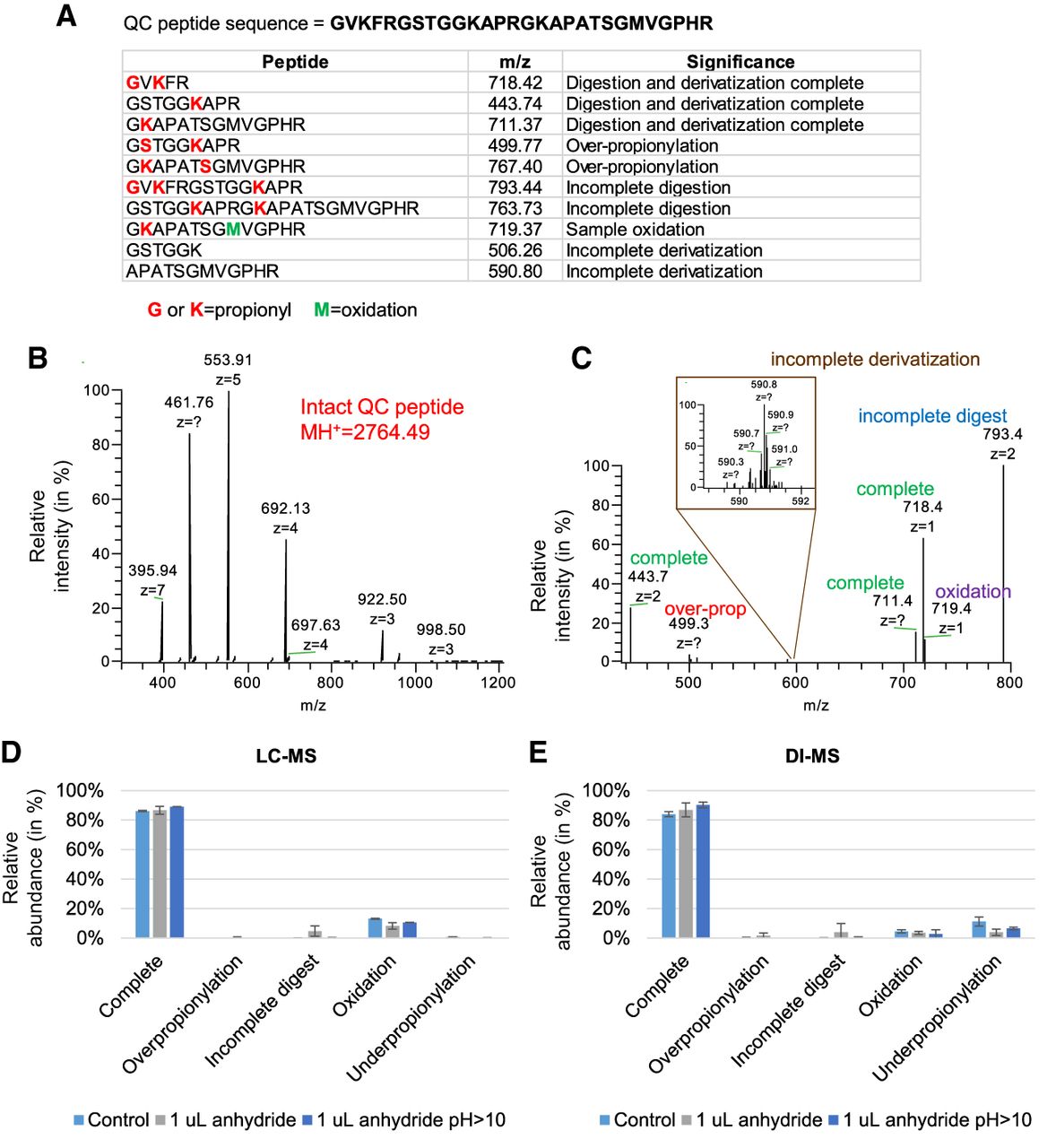
Analysis of the QC peptide. (A) Sequence of the QC peptide (top) and theoretical masses of the products of derivatization and digestion. The indicated m/z was the most intense charge state determined by manual observation of the spectra. (B) Full MS spectrum of the peptide underivatized and undigested. (C) Example of signals detectable after derivatization and digestion. The spectrum was acquired after 5 min digestion (to detect evidence of undigested forms) using a targeted-SIM multiplexed (MSX) scan. (D) Comparison of three sample preparation protocols using the QC peptide detected by LC-MS and (E) DI-MS. Control is defined as 2 h digestion at pH 8 using 5 µL of propionic anhydride; the second protocol used only 1 µL of propionic anhydride, and the third used an excess of ammonium hydroxide to bring the pH > 10. The QC peptide was prepared in a background of endogenous histones purified from mouse stem cells. The error bar represents the standard deviation of three biological replicates.


