
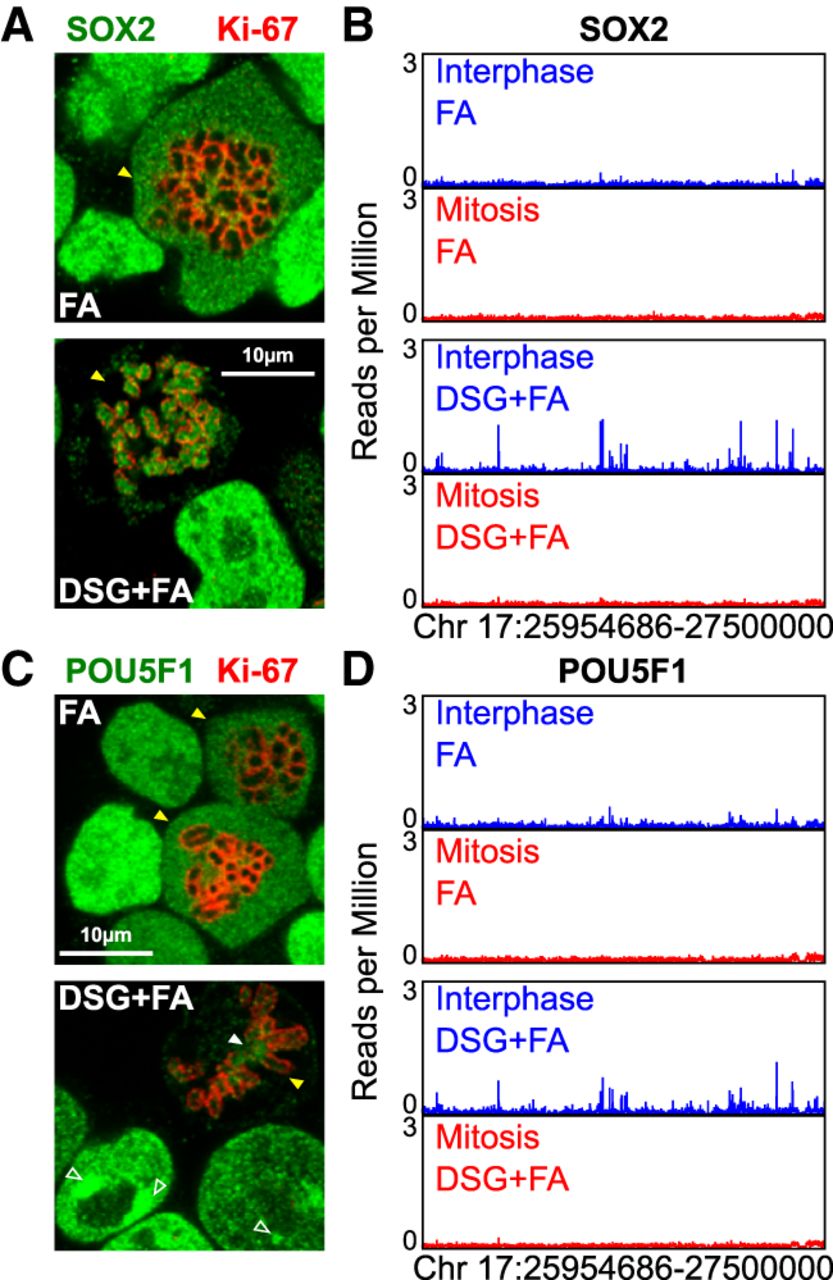
Figure 2.
SOX2 and POU5F1 do not bind at regulatory regions in mitosis. (A) SOX2 immunofluorescence (green), after fixation with either FA (top) or DSG+FA (bottom). The mitotic chromosome periphery is identified by Ki-67 (red). (B) Representative binding profiles of SOX2 presented as in Figure 1D. (C,D) Results of the same analyses described in A and B are shown for POU5F1. Mitotic cells are indicated in A and C with yellow arrowheads. In C, the filled white arrowheads point to the centromeres, the empty white arrowheads to the PCH.


