
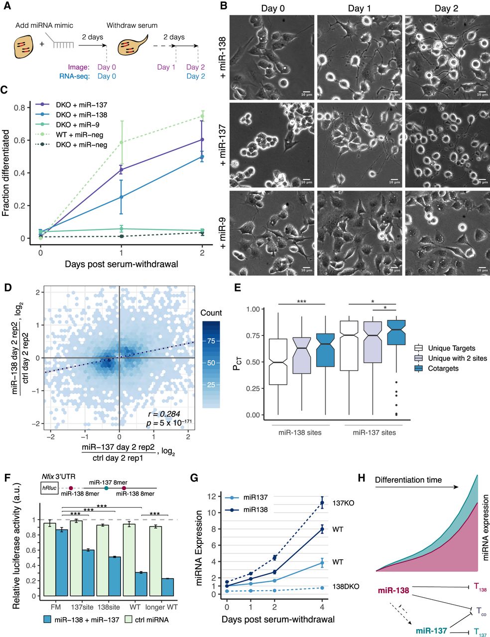
Both miR-138 and cotargeting partner miR-137 can rescue the neurite growth phenotype of mir-138 DKO cells. (A) DKO or WT cells were transfected with a miRNA mimic, and serum was withdrawn 2 d later. Images were taken 2 d after the transfection, before serum withdrawal (day 0), and 1 and 2 d after serum withdrawal (days 1, 2). RNA-seq libraries were prepared from samples 0 and 2 d after serum withdrawal. (B) DKO cells transfected with a miRNA mimic (miR-138, miR137, or miR-9) and imaged at 0, 1, and 2 d after serum withdrawal (20× magnification; scale bar, 10 µm). (C) Quantitation of morphological differentiation at 0, 1, and 2 d after serum withdrawal of WT cells transfected with a negative control miRNA mimic (miR-neg) and of DKO cells transfected with miR-137, miR-138, miR-9, or miR-neg mimic. (D) Hexagonal heatmap of significantly changing genes in DKO cells transfected with miR-138 or miR-137 mimic, normalized to different control replicates transfected with pUC19 DNA (regression line in dotted dark blue; r = 0.28, P = 5 × 10−171). (E) Probability of conserved targeting (PCT) scores of miR-138 and miR-137 sites in targets containing only one target site for that miRNA (white), targets containing two sites for the same miRNA (purple), or cotargets containing one target site for miR-138 and one target site for miR-137 (blue). Wilcoxon rank-sum test: (*) P < 0.05, (****) P < 0.001). (F) Relative luciferase signal (Renilla/firefly) for psiCHECK-2 reporter containing a 500-bp region of the Nfix 3′ UTR with one miR-138 and one miR-137 site (WT) or with a combination of seed site mutations: both miRNA sites mutated in a full mutant (FM), only the miR-138 site mutated (137 site), only the miR-137 site mutated (138 site), or an 800-bp region of the 3′ UTR containing an additional miR-138 site (longer WT). Cotransfection with miR-138 and miR-137, or a control miRNA (cel-miR-67), was normalized to a transfection with no miRNA mimic added. Select comparisons are shown (t-test): (*) P < 0.05, (**) P < 0.01, (***) P < 0.001). (G) Relative expression of miR-137 (light blue) or miR-138 (dark blue) measured by TaqMan qPCR (miR/U6) in WT and 138 DKO or 137 KO cells at 0, 1, 2, and 4 d after serum withdrawal, normalized to WT day 0. (H) Summary of miR-138 and miR-137 regulation during neuronal differentiation, showing repression of individual and shared (Tco) targets, and (indirect) regulatory relationships between the miRNAs. See also Supplemental Figure S3 and Supplemental Table S4.


