
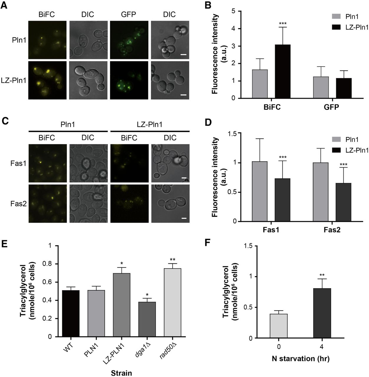
Involvement of Pln1 in lipid metabolism. (A) Representative BiFC and GFP images of cells expressing Pln1 and LZ-Pln1. Scale bars, 2 μm. LZ-Pln1 represents Pln1 tagged with the leucine zipper (LZ) motif. (B) Quantification of the fluorescence intensity of cells in A. Gray and black bars indicate the fluorescence intensity of cells expressing Pln1 and LZ-Pln1, respectively. Error bars, SD. Asterisks indicate significant differences compared with Pln1 (Student's t-test): (***) P < 0.001. (C) Representative BiFC image of cells expressing Pln1- or LZ-Pln1-VN and Fas1- or Fas2-VC. Scale bars, 2 μm. (D) Quantification of the fluorescence intensity of cells in C. Gray and black bars indicate the fluorescence intensity of cells expressing Pln1 and LZ-Pln1, respectively. Quantitative and statistical analysis was performed as described in B. (E) Measurement of TAG contents. TAG was measured as described in the Supplemental Material. Values represent the average of eight independent experiments; error bars, SD. dga1Δ and rad50Δ cells were used as a negative and a positive control, respectively (Oelkers et al. 2002; Kanagavijayan et al. 2016). Asterisks indicate significant differences compared with wild-type cells (Student's t-test): (*) P < 0.05, (**) P < 0.01. (F) TAG contents of cells under nitrogen starvation. The TAG levels in wild-type cells under the normal conditions and nitrogen starvation were measured. Values represent the average of three independent experiments; error bars, SD. Asterisks indicate significant differences compared with the normal conditions (Student's t-test): (**) P < 0.01.


