
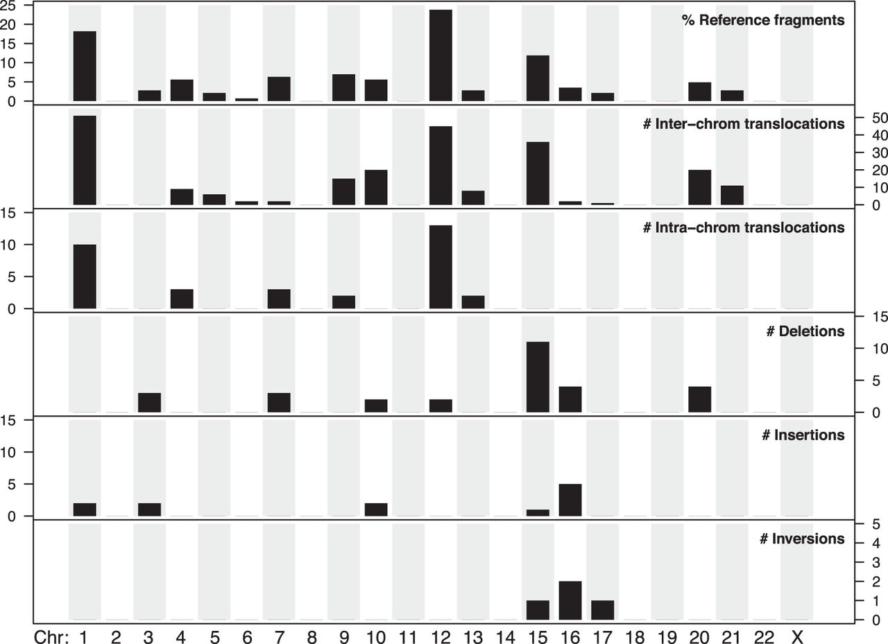
Figure 5.
Chromosomal distribution of donor sequences and five SV types observed in 72 fusion maps. Top panel shows the percentage of reference donor fragments found in the fusion maps belonging to each of the 22 autosomes and Chromosome X. The next five panels show the numbers of each SV event involving the corresponding chromosomes.


