
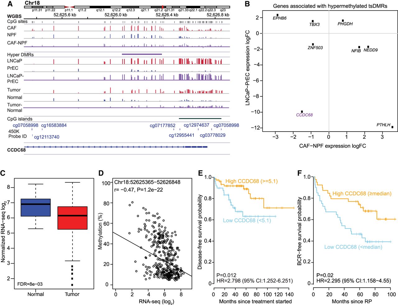
CCDC68 has prognostic value in localized prostate cancer. (A) WGBS data showing consistent hypermethylation of CCDC68 in CAFs versus NPFs (n = 4 pairs), LNCaP versus PrEC, and matched tumor versus normal tissues (n = 4 pairs). The locations of 450K probes are shown in gray. (B) Scatter plot of differentially expressed genes associated with hypermethylated tsDMRs. CCDC68 (purple) is down-regulated in CAFs versus NPFs (n = 4 pairs) and also in LNCaP versus PrEC cells. (C) Box plot showing decreased CCDC68 expression in tumor (n = 392) versus normal (n = 45) tissue samples from TCGA. (D) The relationship between 450K methylation averaged across all probes at the TSS of CCDC68 and gene expression for TCGA tumor specimens. (E,F) Kaplan–Meier curves showing low (blue) CCDC68 expression is associated with poor recurrence-free survival compared to high CCDC68 expression (orange) in Taylor (E) and Glinsky (F) data sets (n = 127 and n = 79 respectively, log-rank test).


