
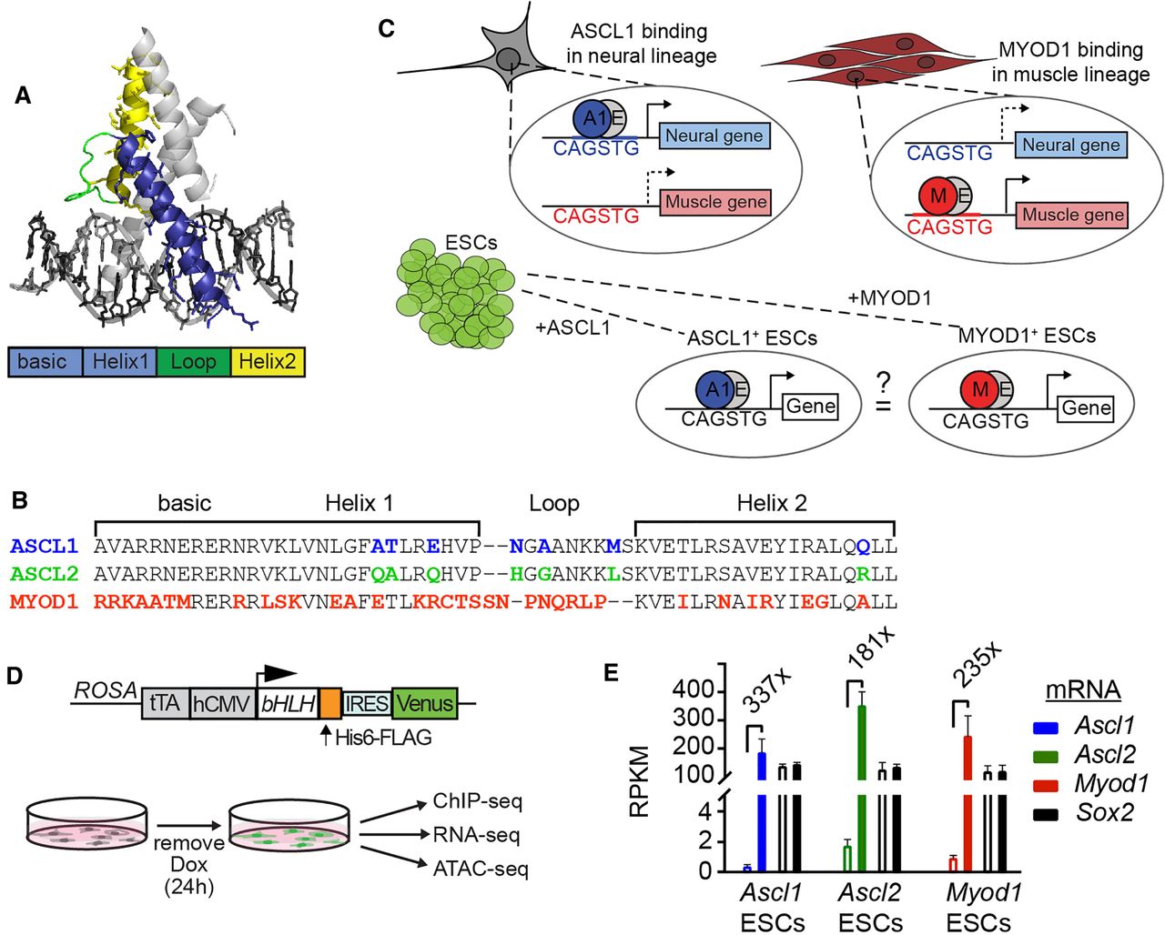
ASCL1, ASCL2, and MYOD1 are related class II bHLH transcription factors. Class II bHLH factors heterodimerize with class I bHLH factors and selectively bind CANNTG Eboxes in vitro (Johnson et al. 1992; Murre et al. 1994). (A) Illustration showing conserved basic helix-loop-helix structural domains modeled on the NEUROD1:E47 crystal structure (Longo et al. 2008). Colored bar shows defined domains and corresponds to the colors on the structure. (B) Comparison of the amino acid sequence of ASCL1, ASCL2, and MYOD1 bHLH domains. The colors represent distinctions within the bHLH domain unique to only one of the proteins compared. (C) Illustration showing the concept tested in this study. Previous work shows that ASCL1 and MYOD1 bind to distinct sites in the lineages where they are expressed, neural and muscle, respectively; however, they recognize the same CAGSTG motif. We ask whether they will bind the same sites or distinct sites when presented with the same chromatin landscape. (D) Diagram of the transgene present in the ESC lines that express a single bHLH factor when cultured in the absence of doxycycline (DOX) (Gossen and Bujard 1992; Guturu et al. 2013). (E) RNA-seq RPKM values from ESCs before and after induction of the individual bHLH factors (Supplemental Table S2). The mean fold change across replicates is shown for each. mRNA levels for an endogenous TF in ESCs, Sox2, are shown for comparison.


