
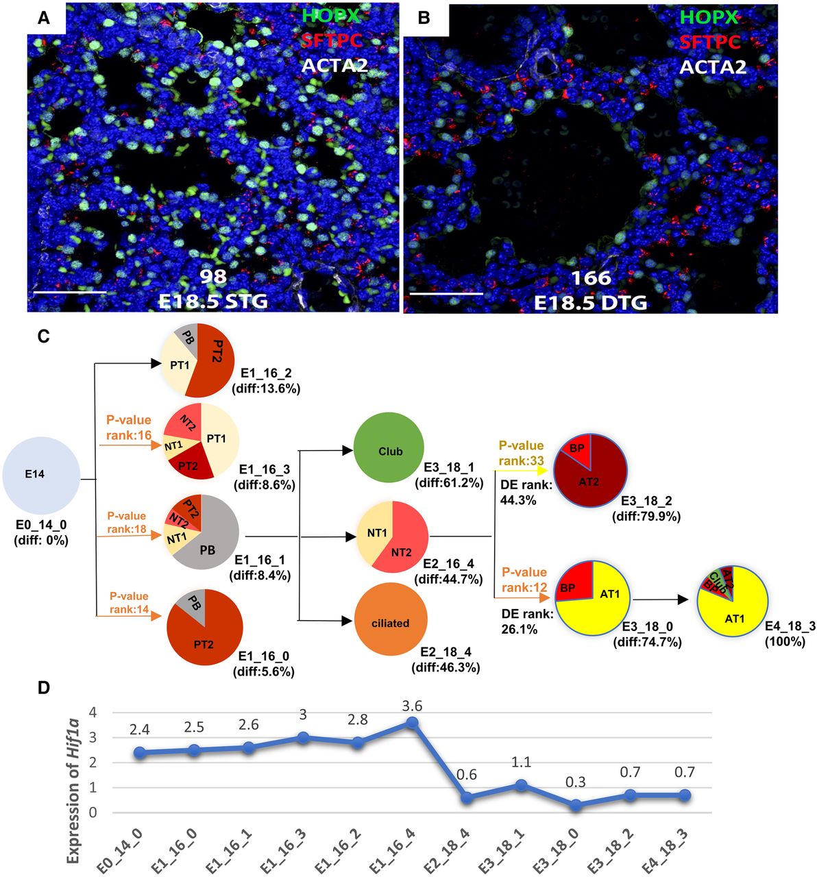
Increased HIF1A activity disrupts sacculation and influences AT1/AT2 cell distribution. (A,B) Experimental results for HIF1A staining. HIF1A (three-point mutant) was expressed under conditional control of SFTPC-rtTA, (otet)7-HIF1A-TPM. Doxycycline was provided to the dam from E12.5 to E18.5. Single transgene (STG) controls, lacking HIF1A-TPM expression (n=3), were compared with double transgenic (DTG) mice expressing HIF1A-TPM under doxycycline control in airway epithelial cells (n=4). Staining of lung tissue for ACTA2 (smooth muscle actin), HOPX (an AT1 cell marker), and SFTPC (proSP-C, and AT2 cell marker) are shown. (C) Model prediction for HIF1A. HIF1A is identified as a top regulator of AT1 cells (ranked as the 12th TF) with a lower, though still significant, impact on AT2 cells (ranking as the 33rd TF). (D) mRNA expression of Hif1a in the different states reconstructed by the model. As predicted by the model, OE of Hif1a influences AT1/AT2 cell distribution with a larger impact on AT1 cells compared with AT2 cells.


