
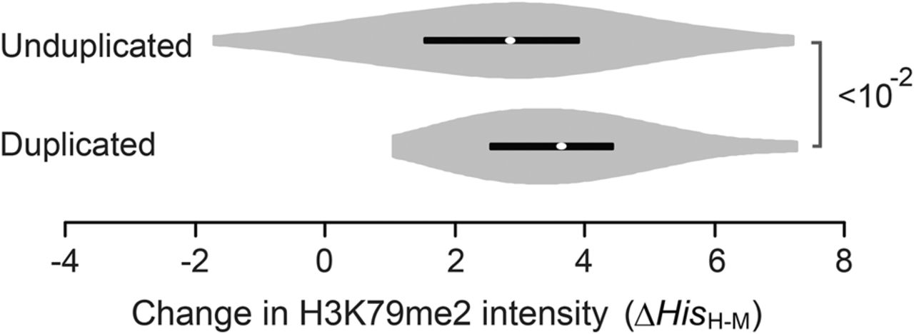
Figure 5.
Violin plots of ΔHisH-M of H3K79me2 at “TSS to TSS+500” that were measured in mammalian liver tissues. Orthologs that underwent duplication events (duplicated) and those that did not (unduplicated) were compared in the mouse lineage. Only orthologs composed of a single copy of human genes with a H3K79me2 signal greater than zero were analyzed. P-values were determined with the Mann-Whitney U test.


