
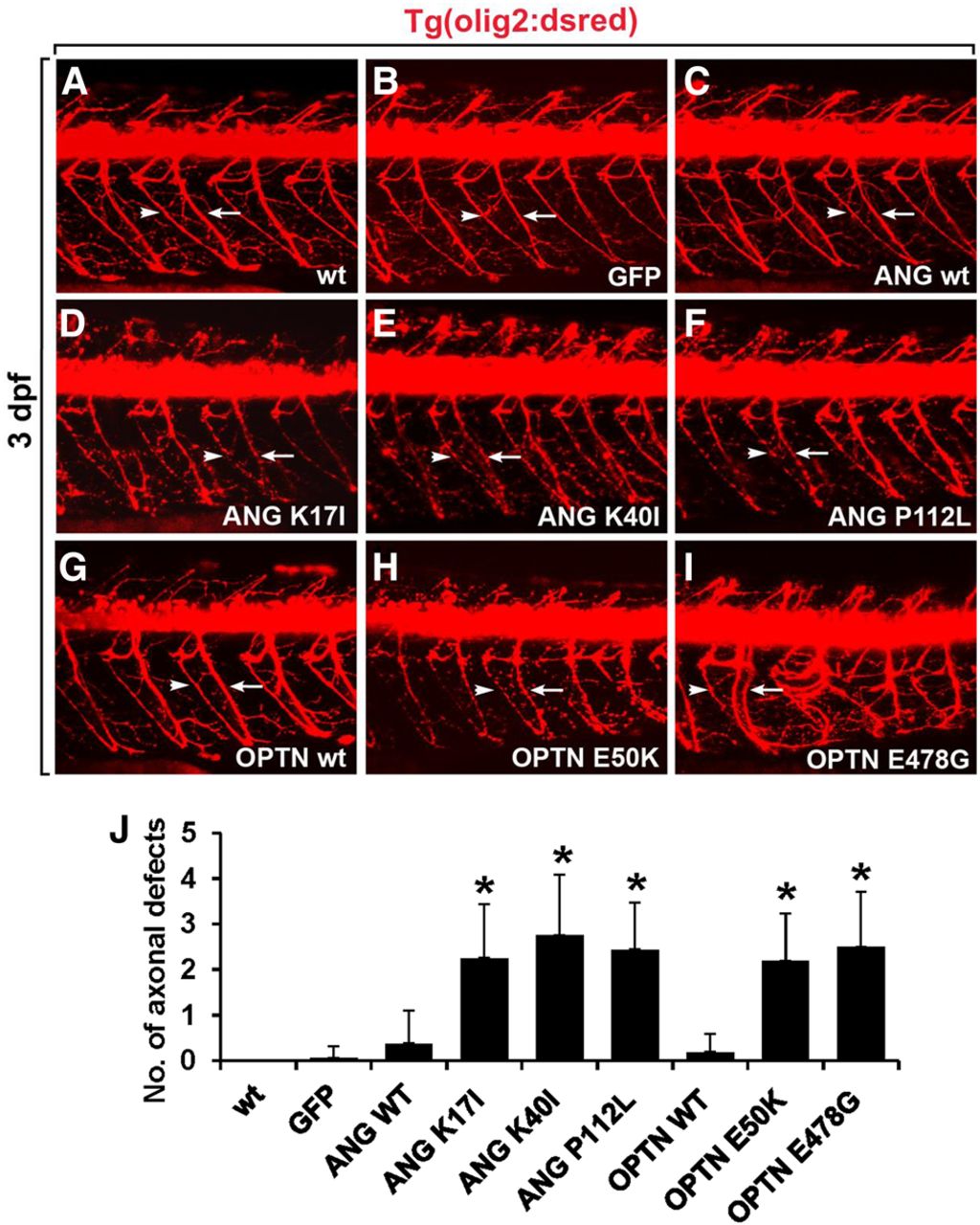
Overexpression of ANG or OPTN mutants causes motor axonopathy in the spinal cord of zebrafish embryo. All panels show lateral views of the spinal cord of Tg(olig2:dsred2) embryos, with anterior to the left and dorsal to the top. Motor axons (arrows) and neuromuscular junctions (NMJs, arrowheads) were detected with DsRed fluorescent protein expression. (A,B) Visualization of motor axons and neuromuscular junctions in the noninjected (A) and egfp mRNA-injected control embryos (B). (C–F) Injection of mRNA for wild-type ANG (C), ANG K17I (D), ANG K40I (E), and ANG P112L (F) mutants in the Tg(olig2:dsred2) embryos. (G–I) Injection of mRNA for wild-type OPTN (G), OPTN E50K (H), and OPTN E478G (I) mutants in Tg(olig2:dsred2) embryos. (J) Statistical analysis of A–I. Axonal defects included axonal swelling and degeneration. Data were obtained from 10 control and 10 mRNA-injected embryos. (*) P < 0.05 versus GFP-expressing control embryos; mean ± SD.


