
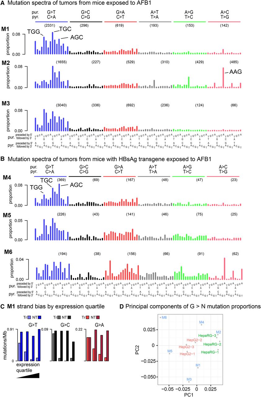
Somatic mutation spectra from HCC-like liver tumors from (A) three AFB1-exposed mice and (B) three AFB1-exposed mice with an HBsAg transgene. The latter have only one-tenth as many mutations as tumors from the AFB1-only mice. Spectra in panels A and B were normalized to the trinucleotide frequencies in the human genome. (C) Extreme transcription-strand bias for all G > N mutations in highly expressed genes in mouse M1. See Supplemental Figure S3 for other mice. Because of low mutation count, transcription-strand bias is evident only in the G > T mutations in the tumors from the AFB1 + HBsAg mice. Transcribed strand (Tr); nontranscribed strand (NT). (D) Principal components analysis (PCA) on G > N mutations in trinucleotide context. Replicates of each of the cell lines cluster together, while the mouse tumors are more dispersed in principal components space. The greater dispersion among the HBsAg tumors (M4, M5, M6) is likely due to higher stochastic variance because of much lower mutation counts combined with greater relative contributions from other mutational processes that arose during tumor development.


