
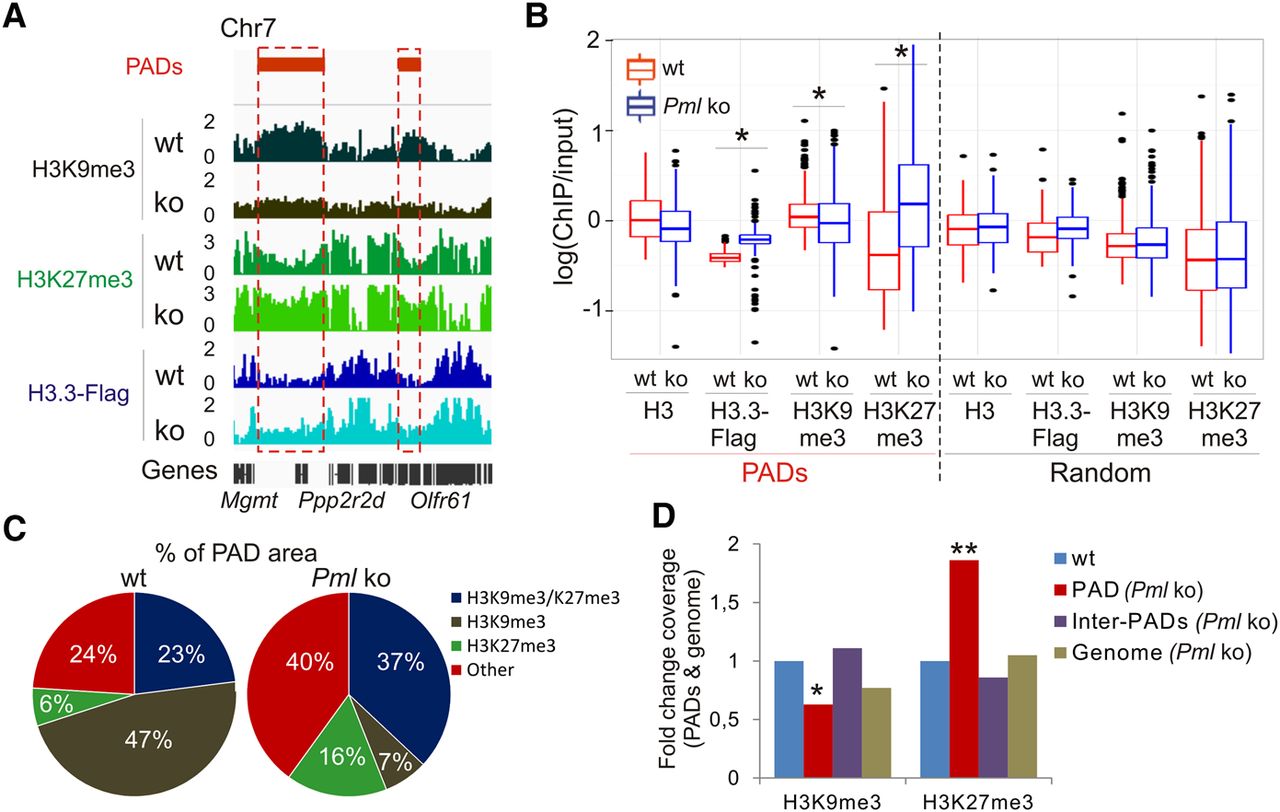
Figure 4.
The heterochromatic state of PADs is remodeled in the absence of PML. (A) Profiles of H3K9me3, H3K27me3, and H3.3-Flag inside and outside PADs in wt and Pml ko MEFs. (B) Median enrichment level of indicated marks in PADs and in randomized PADs. (*) P < 2.2 × 10⁻¹⁶; Wilcoxon tests. (C) Proportions of PAD area enriched in H3K9me3 and H3K27me3. (D) Fold change of H3K9me3 and H3K27me3 coverage in PADs (red bars), between PADs (purple bars), and in the whole genome (brown bars) in Pml ko versus wt cells. Reference (wt) coverage is set to one (blue bars). (*) P < 0.01, (**) P < 0.001 relative to wt; one-sample t-tests.


