
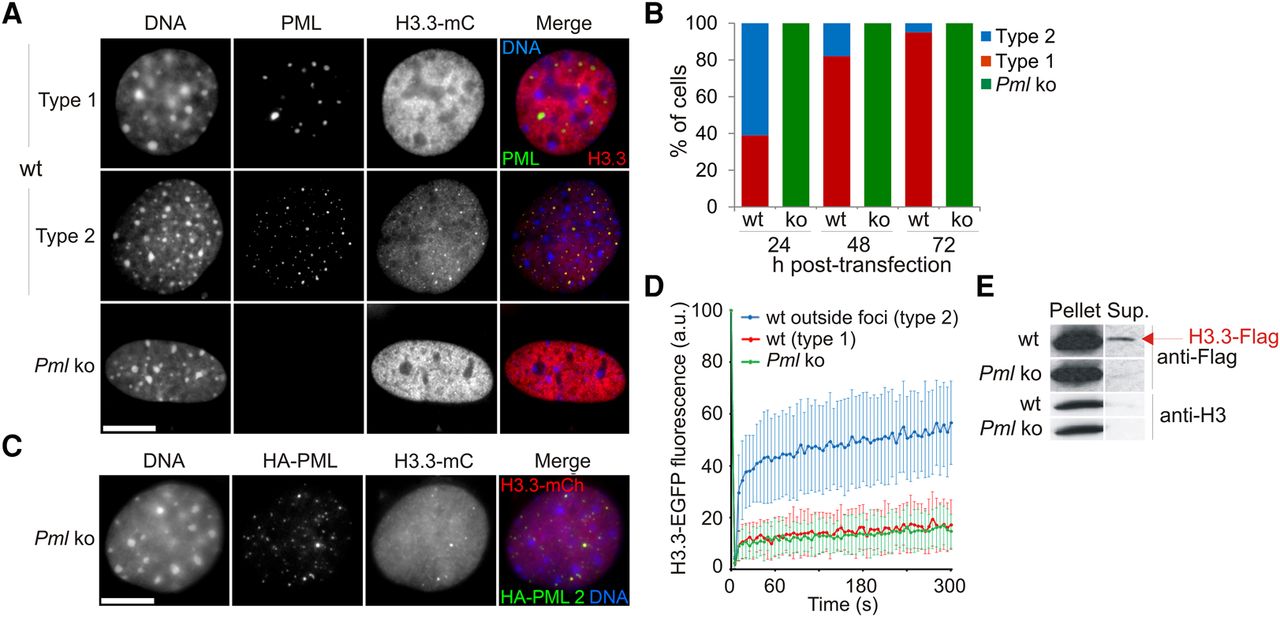
Loss of PML accelerates deposition of epitope-tagged H3.3 into chromatin. (A) Distribution of H3.3-mCherry (mC) in wt and Pml ko MEFs 24 h after H3.3-mC transfection. Cells were labeled using anti-PML antibodies and DNA stained with DAPI. Bar, 10 µm. (B) Proportions of cells of “type 1” and “type 2” over time after H3.3-mC transfection; no “type 2” Pml ko cells are detected at any time point. (C) Expression of HA-PML (full-length isoform PML2) in Pml ko MEFs restores H3.3-mC targeting to PML bodies. Bar, 10 µm. (D) FRAP analysis of H3.3-EGFP in wt and Pml ko cells; mean ± SD for 19–33 cells of each indicated type. See Supplemental Figure 2C for visualization of photobleached areas. (E) Western blot of H3.3-Flag and endogenous H3 in a 1% Triton X-100 soluble (Sup.) and insoluble (Pellet) fraction in wt and Pml ko MEFs.


