
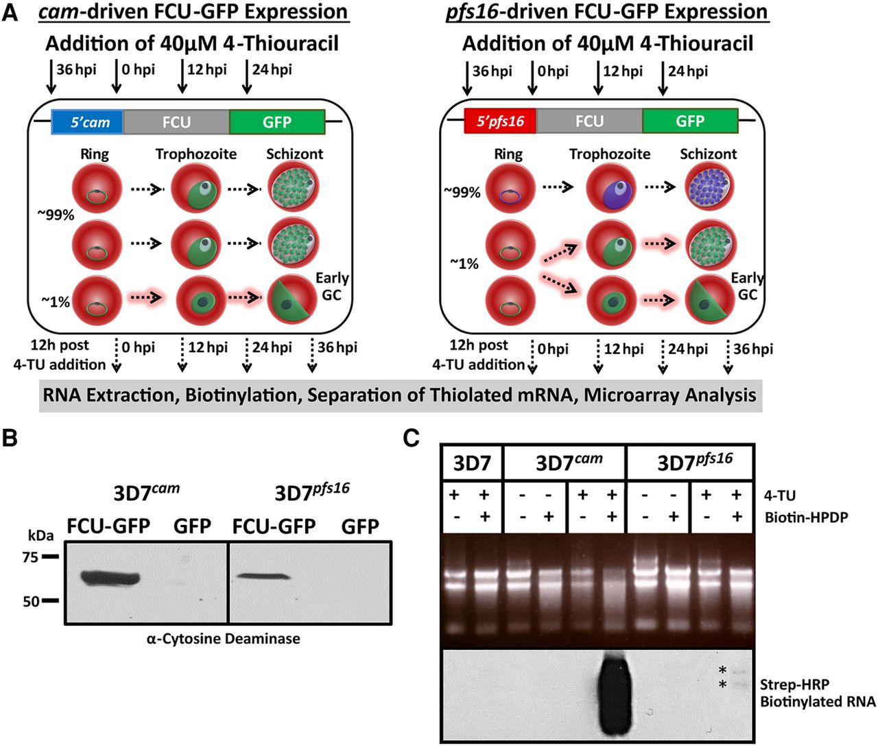
Stage-specific pyrimidine salvage for detection of early gametocyte transcription. To measure mRNA from a subpopulation of cells, we expressed FCU-GFP under the control of the gametocyte-specific promoter pfs16 in the 3D7 parasite line (3D7pfs16). (A) Schematic representation of the experimental design including the timing of 4-TU incubation (black arrows) and RNA extraction (dashed black arrows), plasmids transfected into P. falciparum strains, and a depiction of the highly synchronous cell populations that express cam- and pfs16-fcu-gfp throughout the 48-h IDC. All parasites express FCU-GFP from the constitutive cam promoter (green) regardless of developmental stage (left, 100% GFP+). Only a small proportion (∼1% GFP+) of parasites becoming committed (dashed highlighted arrows) during the IDC, and those that have entered gametocytogenesis in the previous cycle and are sexually committed rings express FCU-GFP from the pfs16 promoter (green), whereas asexual parasites do not (right, ∼99% GFP−). (B) FCU-GFP protein from uninduced asexual cultures of 3D7cam and 3D7pfs16 was detected by Western blot probed with anti-yeast cytosine deaminase. (C) Detection of the subpopulation of FCU-mediated thiol-tagged RNA was carried out by incubating highly synchronized P.f. 3D7, 3D7cam, and 3D7pfs16 in the presence or absence (top) of 40 µM 4-TU for 12 h. Total RNA was extracted, biotinylated, and assayed by Northern blot probed with Streptavidin-HRP (bottom), demonstrating that thiol tagging occurs at a much lower level in 3D7pfs16 (*) than in 3D7cam, representing the minor sexual-stage parasite subpopulation.


