
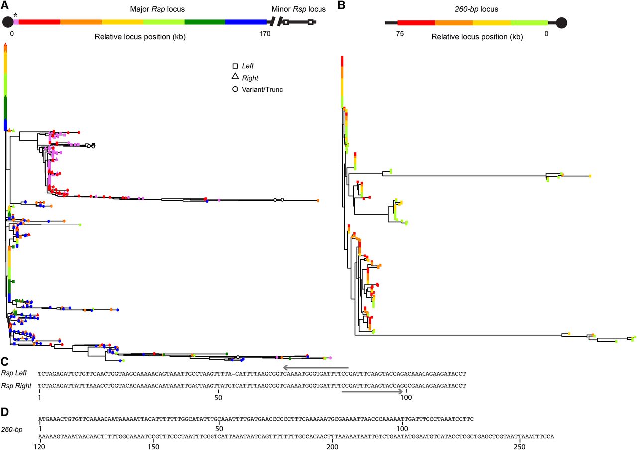
Neighbor-joining tree of complex satDNA monomers. (A) Rsp repeats in the Chromosome 2R locus. Repeats were divided into bins each of which contains one-sixth of the locus, or about 180 repeats/bin. Tip color corresponds to position in the array (red is most centromere-proximal; blue is most distal). The tip symbol indicates if the repeat is Rsp Left (square), Rsp Right (triangle), or variant/truncated (circle). (*) Repeats corresponding to the G2 contig suspected of being centromere-proximal are indicated in pink. Note that these repeats cluster with the repeats on the proximal end of the Rsp contig (red), although it is possible that these are actually distal to the locus. (B) 260-bp repeats in the Chromosome 2L locus. Repeats were divided into bins each of which contains one-fourth of the locus, or about 57 repeats/bin. Tip color corresponds to position in the array (green is most centromere proximal; red is most centromere distal). (C) Aligned consensus Rsp Left and Rsp Right repeat sequences with PCR primers (arrows) used to amplify across Left/Right Rsp dimers. (D) Consensus 260-bp repeat sequence.


