
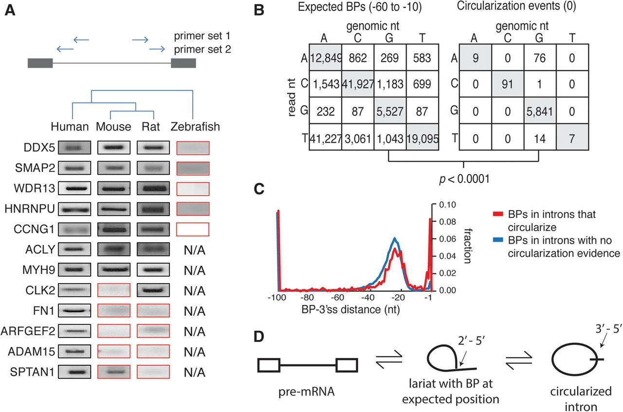
Intron circularization via 3′-5′ linkage is conserved and arises in introns that also contain branchpoints in the expected region. (A) Conservation of the intron circles was assessed by inverted nested RT-PCR in multiple species (primer location indicated by arrows) and Sanger sequencing. The PCR results inconsistent with conserved distal branchpoints are boxed in red. ‘N/A’ indicates certain introns were not tested. (B) Mutational profile of branchpoints from expected region (left) compared with mutational profile from full intron circularization events (right). (C) Location of additional branchpoints in introns which circularize (red) was compared to introns with no observed evidence for circularization (blue). (D) Model for circles implicates a 3′-5′ circular linkage arising from a conventional lariat.


