
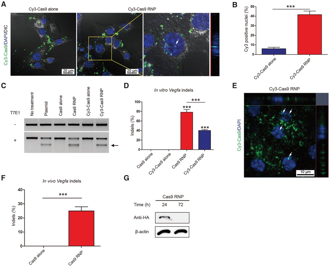
In vitro and in vivo delivery of Cy3-labeled Cas9 RNP. (A) Localization of Cy3 dye in NIH3T3 cells transfected with Cy3-labeled Cas9 RNP or Cy3-labeled Cas9 alone (as a control) at 24 h post-transfection. White arrow indicates nuclear colocalization of Cy3 dye. The z-axis image on the right shows that Cy3-Cas9 is localized inside the nucleus. (B) Proportion of Cy3 positive nuclei in total DAPI positive nuclei at 24 h post-transfection. Error bars indicate SEM (n = 3). Student's t-test: (***) P < 0.001. (C) Vegfa-specific Cas9 RNP-mediated mutations in NIH3T3 cells detected by the T7E1 assay. The arrow indicates the expected position of DNA bands cleaved by T7E1. (D) Mutation frequencies were measured using targeted deep sequencing. Error bars are SEM (n = 3). One-way ANOVA and Tukey post-hoc tests: (***) P < 0.001. (E) Representative RPE flat-mount at day 3 post-injection of Cy3-labeled Cas9 RNP into mouse eye. White arrows indicate nuclear colocalization of Cy3 dye. (F) Frequencies of indels induced in vivo determined using genomic DNA isolated from the retinal pigment epithelium (RPE). Indels were analyzed by deep sequencing at day 3 post-injection. Error bars are SEM (n = 5). Student's t-test: (***) P < 0.001. (G) Representative Western blot analysis to measure the level of Cas9 protein in the RPE/choroid/scleral complex 24 and 72 h after injection (n = 4).


