
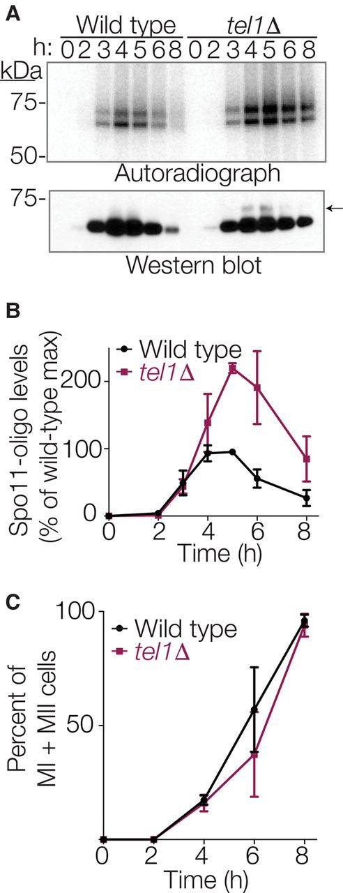
Elevated levels of Spo11-oligo complexes in tel1Δ mutants. (A) Representative time course. Autoradiograph of SDS-PAGE gel (top) shows radiolabeled Spo11-oligo complexes at the indicated times in sporulation medium. Anti-protein A Western blot (bottom) monitors total Spo11. Note that most signal in the Western blot is free Spo11, i.e., not bound to oligos. The arrow indicates the small amount of Spo11 protein that comigrates with slower migrating Spo11-oligo species, visible in the tel1Δ sample because of increased DSB numbers. (B) Quantification of radiolabeled Spo11-oligo complexes in tel1Δ cells, relative to wild-type cultures collected in parallel. Mean ± SD for three pairs of cultures is shown. (C) Meiotic progression (the percentage of cells completing one or both divisions). Mean ± SD for the same cultures as in B; ≥100 cells counted per time point.


