
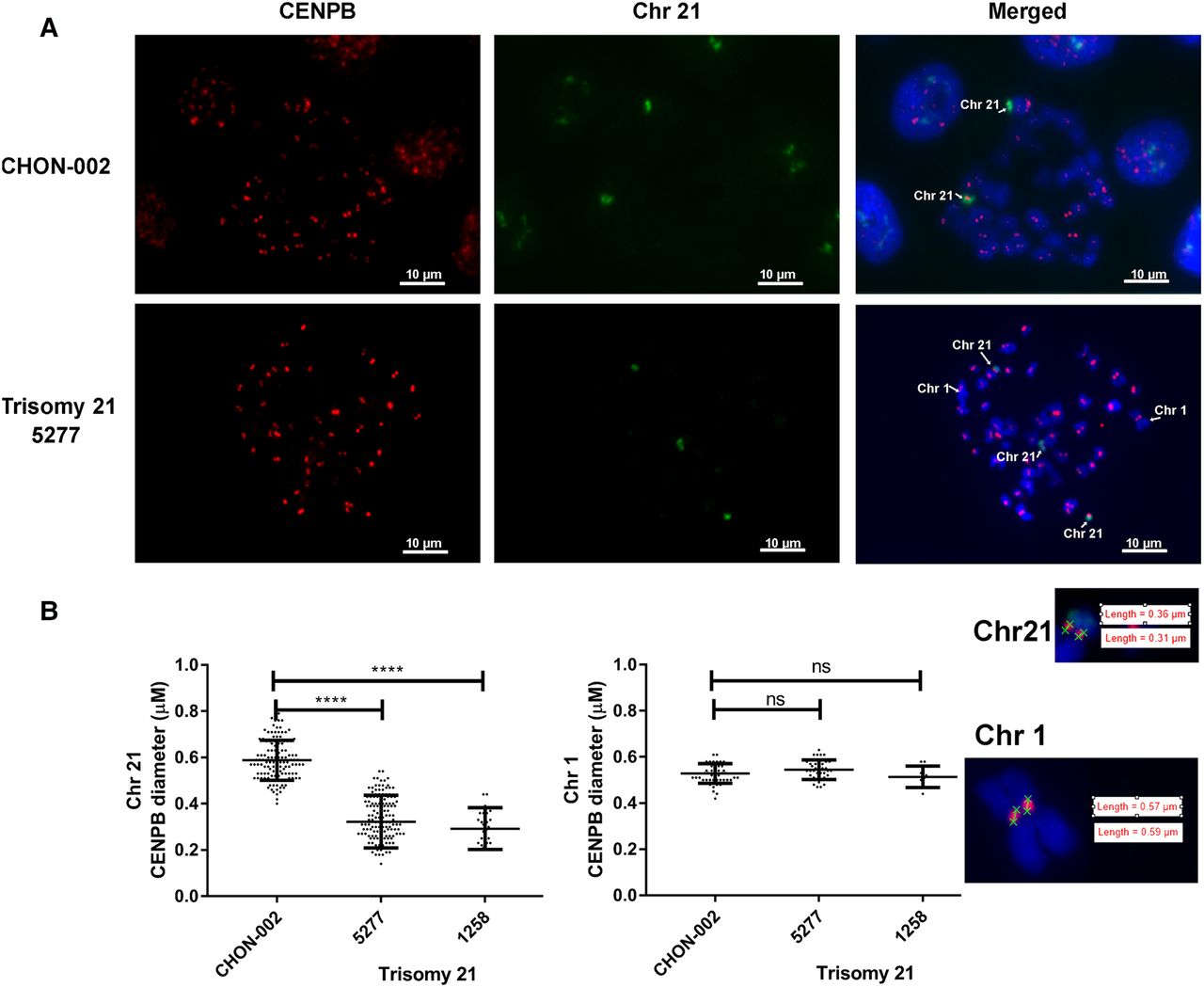
Reduced CENPB binding to the centromere of Chr 21 in trisomy 21 cells. IF-FISH analysis to determine the binding of CENPB (IF) on Chr 21. (A) IF-FISH analysis of CENPB binding (red) on Chr 21 (identified by FISH, green) or Chr 1 (identified by large size) in chromosomal spreads stained with DAPI (blue). Karyotypically normal CHON-002 cells and trisomy 21 5277 cells are shown. (B) The diameter of CENPB binding sites along Chr 21 was measured using the NIS-Elements software in a NIKON microscope in CHON-002 cells and trisomy 21 cells isolated from Subjects A 1258 and B 5277. A statistically significant difference in CENPB binding along Chr 21 was found between the karyotypically normal cell line CHON-002 (n = 126) and the trisomy 21 cells lines 5277 (n = 141) or 1258 (n = 25) (P < 0.0001). No significant differences were found in CENPB binding in Chr 1. The insets show examples of the measurement of CENPB staining in Chromosomes 1 and 21 in trisomy 21 cells.


