
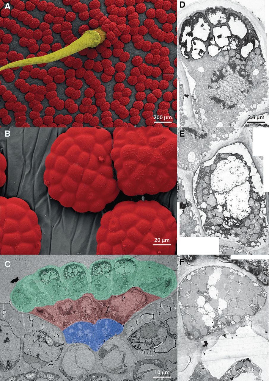
Gland functional morphology. (A) REM false-colored micrograph showing a trigger hair (yellow) surrounded by multiple glands (red) on the inner surface of a Dionaea trap. (B) Close-up of glands (from A). (C–F) TEM micrographs of gland sections. Structural organization of a gland (C) consisting of three functional layers (L1–L3): outer layer L1 (green), inner layer L2 (brown), and endodermoid layer L3 (blue). Secretory cell of the L1 outer layer (D) characterized by large vacuoles and the presence of rough endoplasmic reticulum. Cell of the inner L2 layer (E) exhibiting numerous plasma membrane invaginations. Beta-oxidation probably occurs in L2 cells, which contain big central vacuoles, numerous peroxisomes, and a remarkable number of mitochondria (see also Supplemental Fig. S2). This implies the existence of energy demanding, metabolically active, processes in L2 cells. Endodermoid (stalk) cell (F) comprising layer L3 harboring plenty of oleosomes.


