
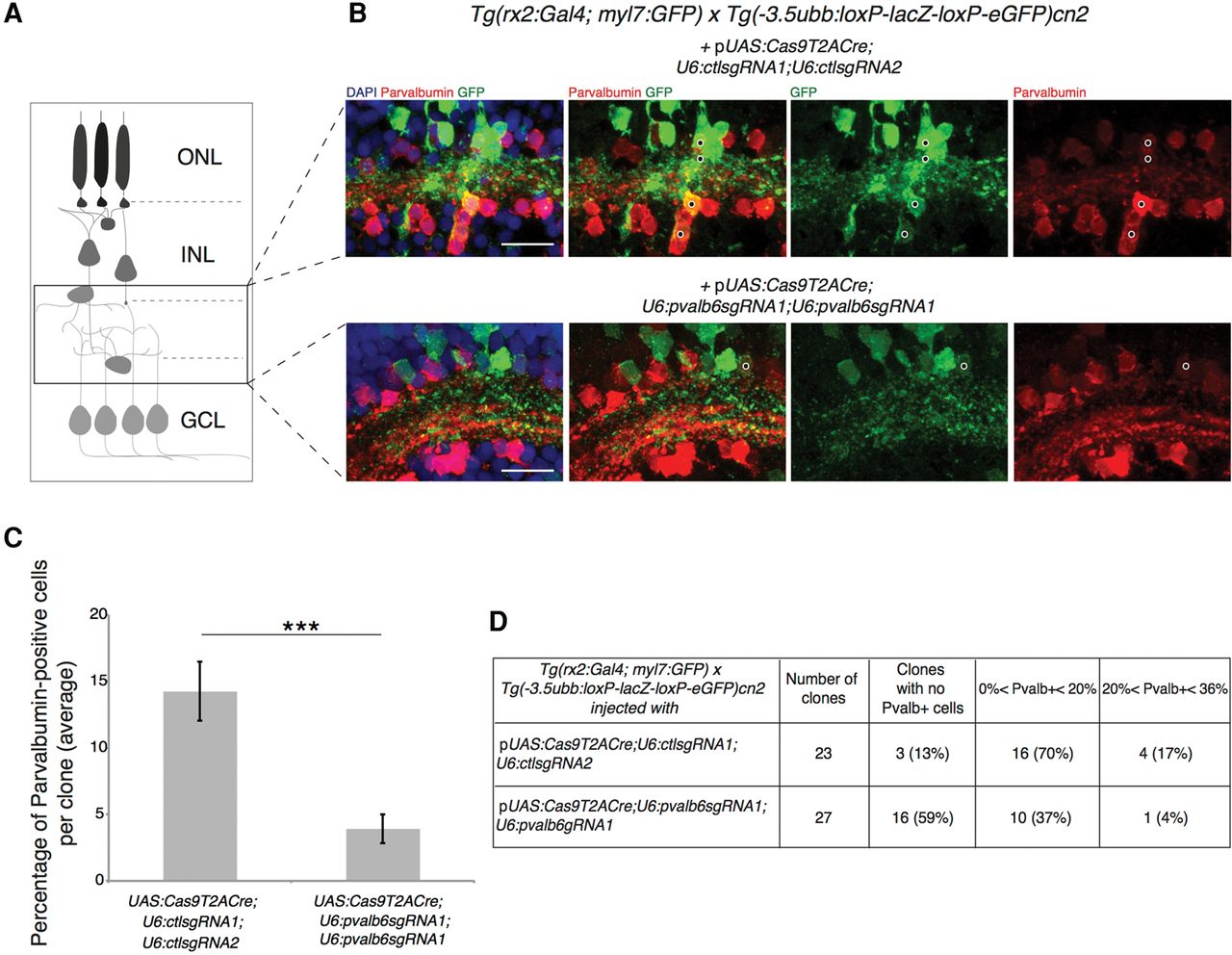
Targeting of the pvalb6 locus leads to loss of Pvalb expression in ACs. (A) Schematic of a clone of cells deriving from a single RPC. The black frame defines the position of the amacrine cell population. (ONL) Outer nuclear layer, (INL) inner nuclear layer, (GCL) ganglion cell layer. (B) GFP-positive Cas9-expressing cells in the retinal section of a 5 dpf double transgenic Tg(rx2:Gal4; myl7:GFP) × Tg (-3.5ubb:loxP-lacZ-loxP-eGFP)cn2 embryos transiently expressing the 2C-Cas9 vector containing control sgRNAs (upper panel) or pvalb6-specific sgRNAs (lower panel). The same sgRNA target sequence was inserted downstream from each U6 promoter (U6:pvalb6sgRNA1). The number of Pvalb- and GFP-double-positive cells (black dots) is reduced if pvalb6 is targeted compared to control. Scale bar = 50 µm. (C) Quantification of the percentage of Pvalb-positive cells per GFP-positive clone. Data are represented as mean ± SEM. (***) P-value < 0.001 following Wilcoxon Mann–Whitney test. (D) Table showing the percentage of Pvalb-positive cells per GFP-positive clone in the retinal sections of double transgenic Tg(rx2:Gal4; myl7:GFP) × Tg(-3.5ubb:loxP-lacZ-loxP-eGFP)cn2 larvae microinjected with the 2C-Cas9 plasmid containing control sgRNAs (first row) or pvalb6-targeting sgRNAs (second row).


