
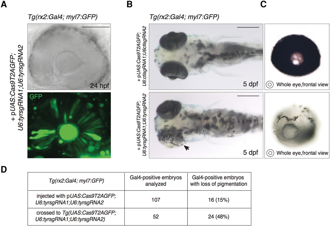
Tissue-specific disruption of the tyrosinase locus induced by the expression of the UAS-based vector in the retinal progenitor cells. (A) Transmitted light and confocal imaging of eye of a 24 hpf Tg(rx2:Gal4;myl7:GFP) embryo injected with pUAS:Cas9T2AGFP;U6:tyrsgRNA1;U6:tyrsgRNA2 together with Tol2 mRNA. GFP fluorescence indicates Gal4-driven expression of the Cas9 enzyme in the retinal progenitor cells (RPCs) giving rise to the retinal pigmented epithelium (RPE) and to the neural retina. Scale bar = 100 µm. (B) (Lower panel) Specific loss of pigmentation in the RPE of a 5 dpf zebrafish embryo induced by injection of pUAS:Cas9T2AGFP;U6:tyrsgRNA1;U6:tyrsgRNA2 in Tg(rx2:Gal4; myl7:GFP) at one-cell stage. Arrowhead indicates the eye with lost pigmentation. (Upper panel) Control embryo injected with the same vector containing a control sgRNA sequence (ctlsgRNA). Scale bar = 300 µm. (C) Frontal view of eyes with absent pigmentation (lower panel) and wild-type (upper panel) explanted from the larvae shown in B. (D) Table showing the percentage of Gal4-positive larvae displaying loss of pigmentation in the RPE. (First row) Injection of the pUAS:Cas9T2AGFP;U6:tyrsgRNA1;U6:tyrsgRNA2 into one-cell-stage Tg(rx2:Gal4; myl7:GFP) embryos. (Second row) Cross of Tg(rx2:Gal4; myl7:GFP) fish with Tg(UAS:Cas9T2AGFP;U6:tyrsgRNA1;U6:tyrsgRNA2).


