
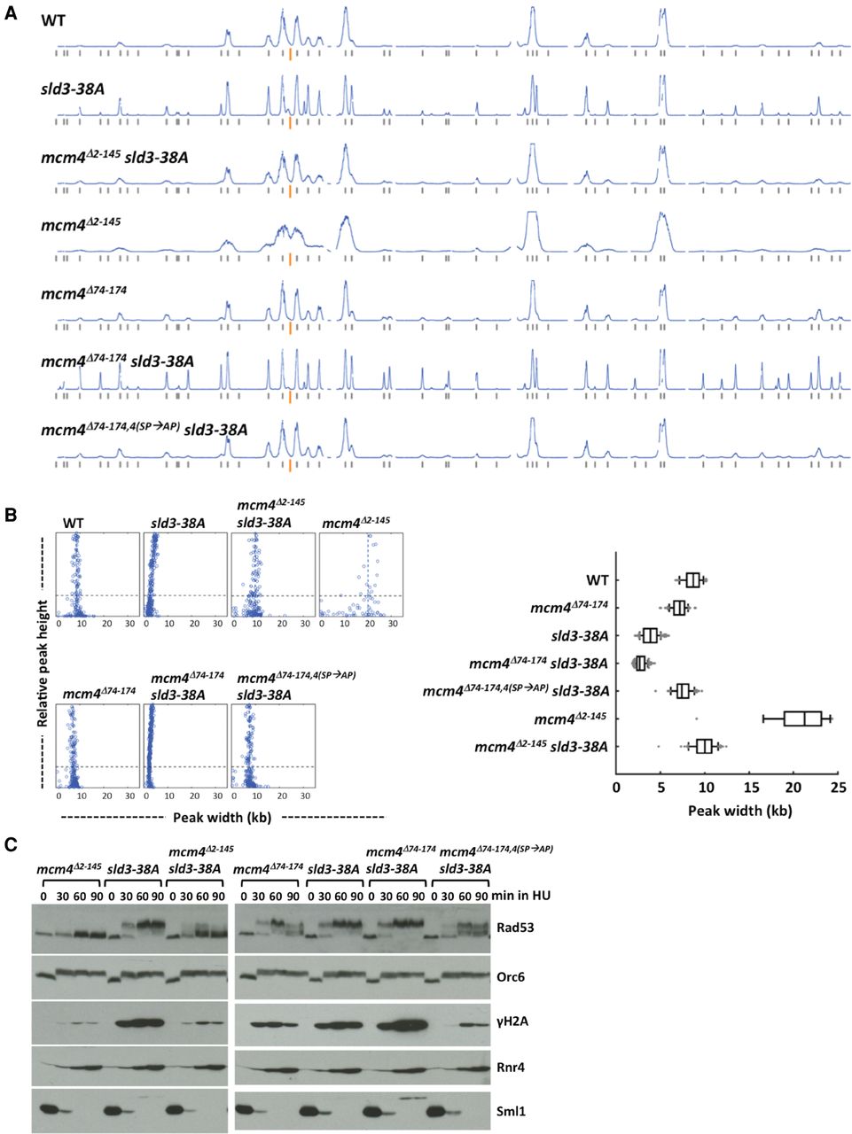
Figure 4.
Effects of sld3-38A and the Mcm4 NSD distal segment on fork progression in HU. (A,B) Yeast cells were synchronized in G1 phase and released into YPD containing 0.2 M HU and 0.5 mM EdU. (A) Replication profiles of Chromosome IV for the indicated yeast strains. (B) Distribution of fork progression from origins shown as individual width–height plots and box graph, excluding peaks with heights <30% of the maximal height scale. (C) Cells from the indicated strains were synchronized in G1, released into 0.2 M HU, and collected at the indicated time points. Protein samples were analyzed as in Figure 2C.


