
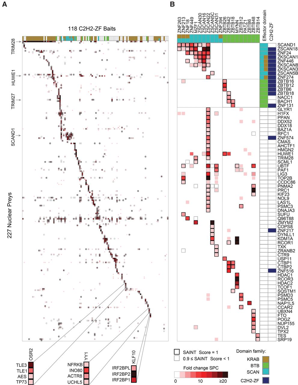
Nuclear protein interactions with C2H2-ZF proteins. AP-MS results for 118 DNA-binding C2H2-ZF proteins. (A) Heat map of PPIs between 118 C2H2-ZF baits and 227 nuclear prey proteins. The fill color represents the fold change spectral counts while the frame color indicates the SAINT score. Colors on top of the heat map represent the effector domain type of the bait proteins. Supplemental Figure S3 contains a version of the heat map with both axes fully labeled. (B) Detailed interactions of all SCAN- and BTB-containing bait proteins. Prey proteins are sorted by their domain type. Colors on top and at the right hand side of the heat map represent the domain types of bait and prey proteins. See also Supplemental Figures S3–S5, Supplemental Tables S5–S9.


