
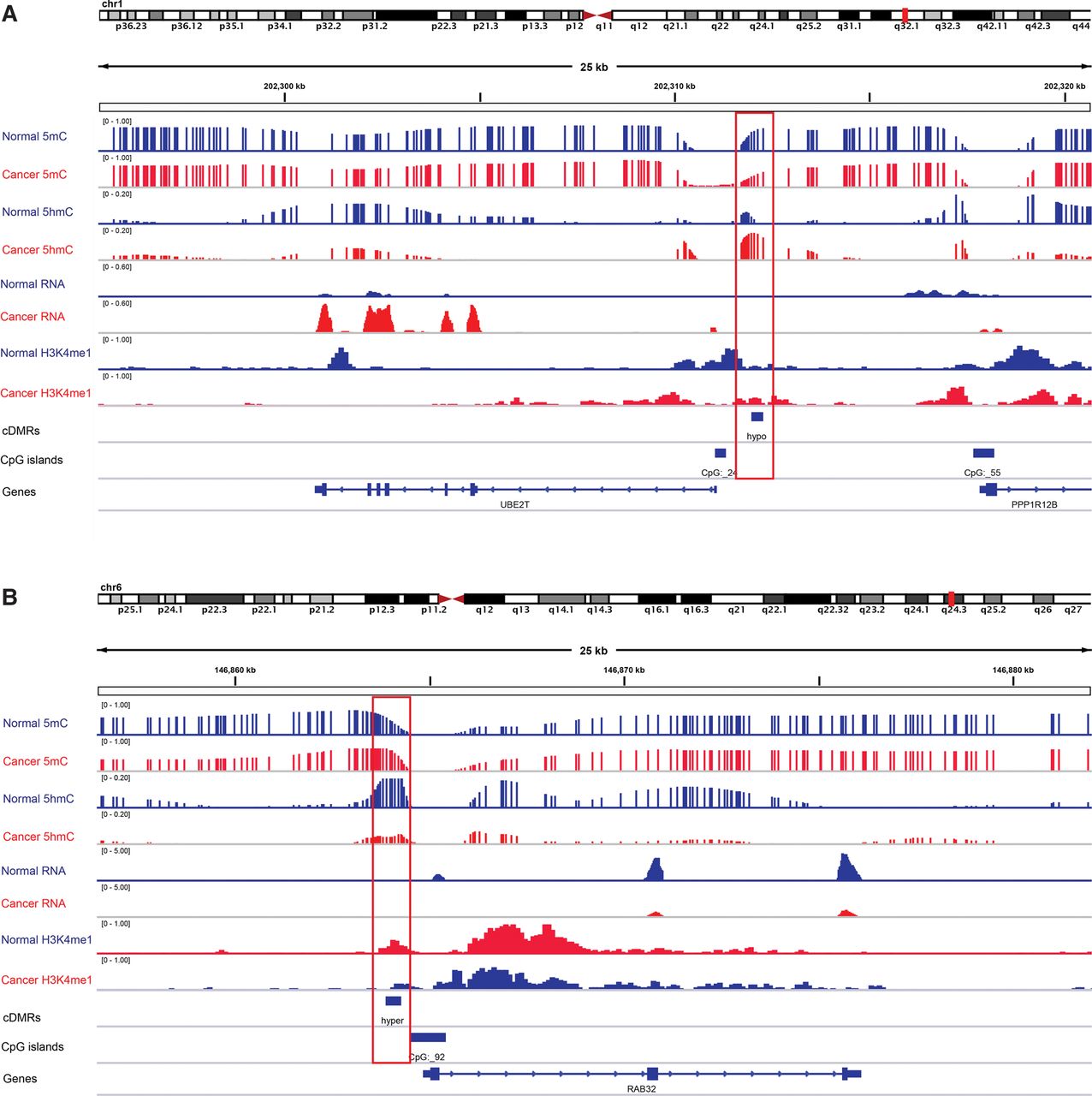
Figure 5.
Two examples of liver c-DMRs that showed 5mC changes were regulated through 5hmC and associated with gene expression changes. 5mC, 5hmC, and gene expression level of both liver normal (blue tracks) and cancer (red tracks) were displayed. Red boxes indicate the location of c-DMRs. (A) A hypomethylated liver c-DMR located in CpG island shores near the promoter of the UBE2T gene. (B) A hypermethylated c-DMR located in CpG island shores near the promoter of the RAB32 gene.


