
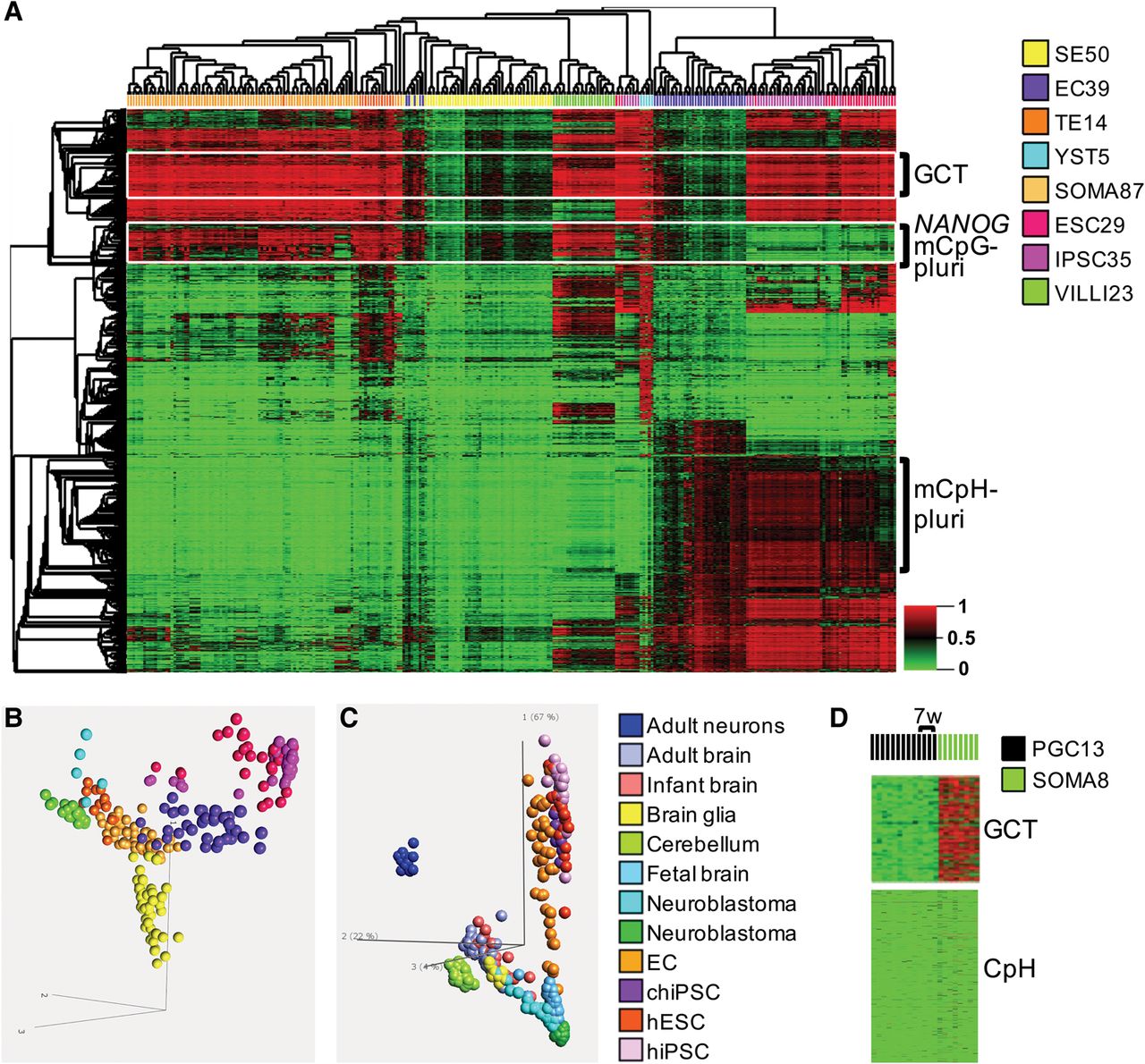
Differentially methylated targets (DMTs) in TGCT subtypes. (A) The methylome profiles of the various TGCT histological subtypes recapitulate embryonic and extra-embryonic epigenomic differentiation. NSE multi-subtype DMTs (selected blinded to reference tissue profiles; see Methods) (see Supplemental Table S3) were used in hierarchical clustering of TGCT and reference GEO database samples representative of pluripotent (ESC), induced pluripotent (iPSC), somatic (SOMA), and extra-embryonic/trophoblastic lineage (placenta tissues and trophoblast cultures). Coclustering emerges for EC with ESC/iPSC, TE with somatic samples, and prominent CpG methylation of YST. The GCT cluster comprises 161 CpG targets uniquely hypomethylated in ECs among NSEs and references, and maps to 76 genes with significantly up-regulated expression in embryonal carcinoma. The “NANOG/PLURI” 141 target module maps to 76 genes, is cohypomethylated in EC/ESC, and includes NANOG, DNMT3B, BCOR, SMAD3, POU5F1, EYA1, FOXH1, HOXB1, HOXB3, LRP4, PBX1, SALL4, TFAP2A, TRIM71, and DPPA4, among others. A striking ESC-like CpH (non-CpG) hypermethylation process was identified in ECs, also present in iPSCs. (B) PCA plot further highlights TGCT-subtype/embryonic-lineage convergence. Color legend at right of A. (C) Pluripotency- versus neuron-signifying CpH methylation. PCA using 473 CpH target module (Methods) (Supplemental Table S3), including ECs and multiple different test groups indicated in color legend at right. PCA shows orthogonality of neuronal versus pluripotency methyl-CpH axes: ECs, and human (hiPSCs), and chimp (chiPSCs) track along the ESC/pluripotency axis, while post-natal cortex and cerebellum lay on the neuronal axis; minimal CpH reprogramming seen in neuroblastoma cell lines (green) and tissues (light blue). (D) Fetal somatic tissues and PGCs as early as 7 wk are erased of CpH methylation, while benign fetal somatic tissue, but not PGCs, maintains methylation at the GCT targets erased in ECs. The numbers of specimens investigated are indicated in the sample color legend. For abbreviations used, see text.


