
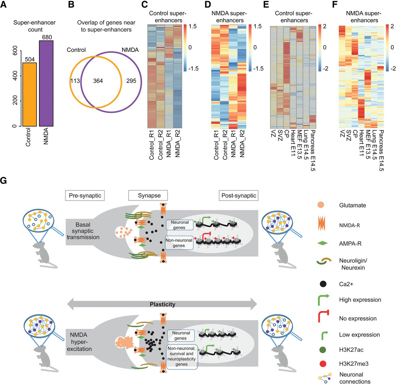
Neuronal activity results in a global reorganization of super-enhancers at key responsive genes. (A) Bar plot showing the number of super-enhancers in the control and NMDA-treated neurons. (B) Venn diagram showing the overlap of genes near control and NMDA super-enhancer. (C,D) Expression of genes near control (C) and NDMA (D) nonpromoter super-enhancers in control- and NDMA-treated neurons (enlarged heat maps along with gene names are provided as Supplemental Fig. S9). (E,F) Heat map showing expression of the same genes as in C and D in tissues from three germ layers. (G) Schematic representation of a model showing epigenome and transcriptome changes in response to sustained NMDA receptor activity. Neurons exhibiting basal synaptic activity express neuronal genes that are marked by an active chromatin state while non-neuronal genes are kept silent in heterochromatin. A prolonged NMDA activity results in a dramatic epigenetic reprogramming, including at many distal regulatory elements and super-enhancers, resulting in the down-regulation of neuronal genes (loss of H3K27ac) and activation of neuronal plasticity, neuroprotective, and several non-neuronal genes (gain of H3K27ac).


