
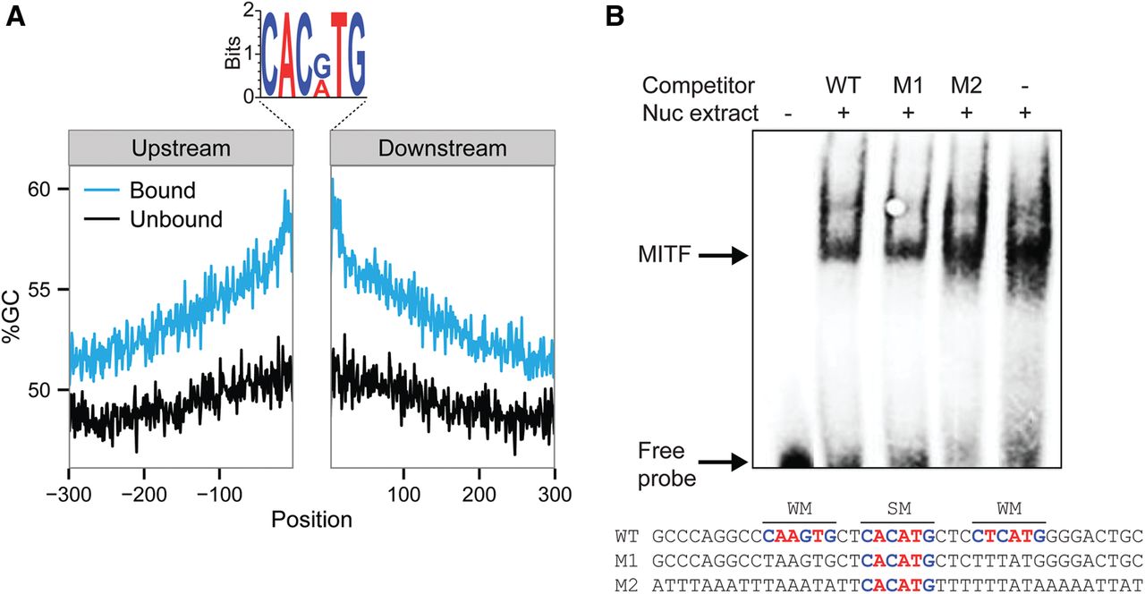
MITF binds to sequences showing overall high similarity to the E-box motif. (A) GC content (%GC) upstream of and downstream from motifs in sequences that are bound (blue) and unbound (black) by MITF. Logo of the MITF bound motifs are shown above: A and T bases are colored in red; G and C bases in blue. (B) EMSA competition assay with probe corresponding to the WT MITF binding region of the human TRPM1 promoter (WT), probe corresponding to the WT with interruption of the two weak MITF motifs (M1), and probe corresponding to the WT with replacement of all G/C by A/T base pair (M2). Highly expressing MITF melanoma cell (WM3682) nuclear extracts were used as a source of MITF (represented in the Nuc extract row above). A biotinylated WT probe was used for the analyses. WT or mutated unlabeled probes as described above were used in the competition analyses. MITF binding probes and free probes are marked with arrows. Probe sequences are shown below, where SM indicates strong motif and WM indicates weak motifs.


