
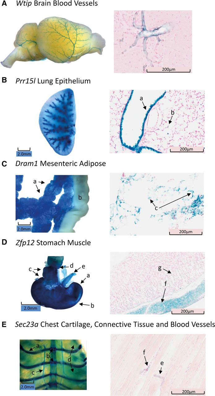
(A) Wtip olfactory bulb, cerebral, and cerebellar blood vessels stain for LacZ in lateral brain whole mount (WM, left), and frozen section (FS, right) reveals staining of a branching cerebral blood vessel. In the Wtip brain, only blood vessels stain for LacZ. (B) Prr15l staining of lung in WM (left) and FS (right). In WM, the major airways clearly stain, but there is also a diffuse staining of the parenchymal tissue. In the FS image, the intense staining of the bronchiole epithelium (a), and the weaker punctate pattern of staining in the walls of the alveoli (b) are indicated. (C, left) Dram1 staining of mesenteric adipose tissue by WM showing diffuse uniform staining (a), while the ileum does not stain (b). (Right) FS of Dram1 mesenteric adipose tissue shows staining is restricted to the stromal, intercellular space (c). (D, left) Zfp12 WM staining of fundus (a), body (b), and antrum (c) of the stomach. WM staining of the pyloris and duodenum (d) and weak staining of the esophagus (e) are similar to that found in WT mice. (Right) FS indicates that the majority of Zfp12 LacZ staining is in the smooth muscle layer (f), although there is weak, scattered staining in the glandular epithelium (g). (E, left) Sec23a WM staining of cartilaginous ribs (a), intersternal plate cartilage (b), blood vessels (c), and a weak striated pattern of staining of the chest muscle (d). (Right) FS of striated Sec23a muscle indicates staining of connective tissue and/or blood vessels (e,f) between muscle fibers.


