
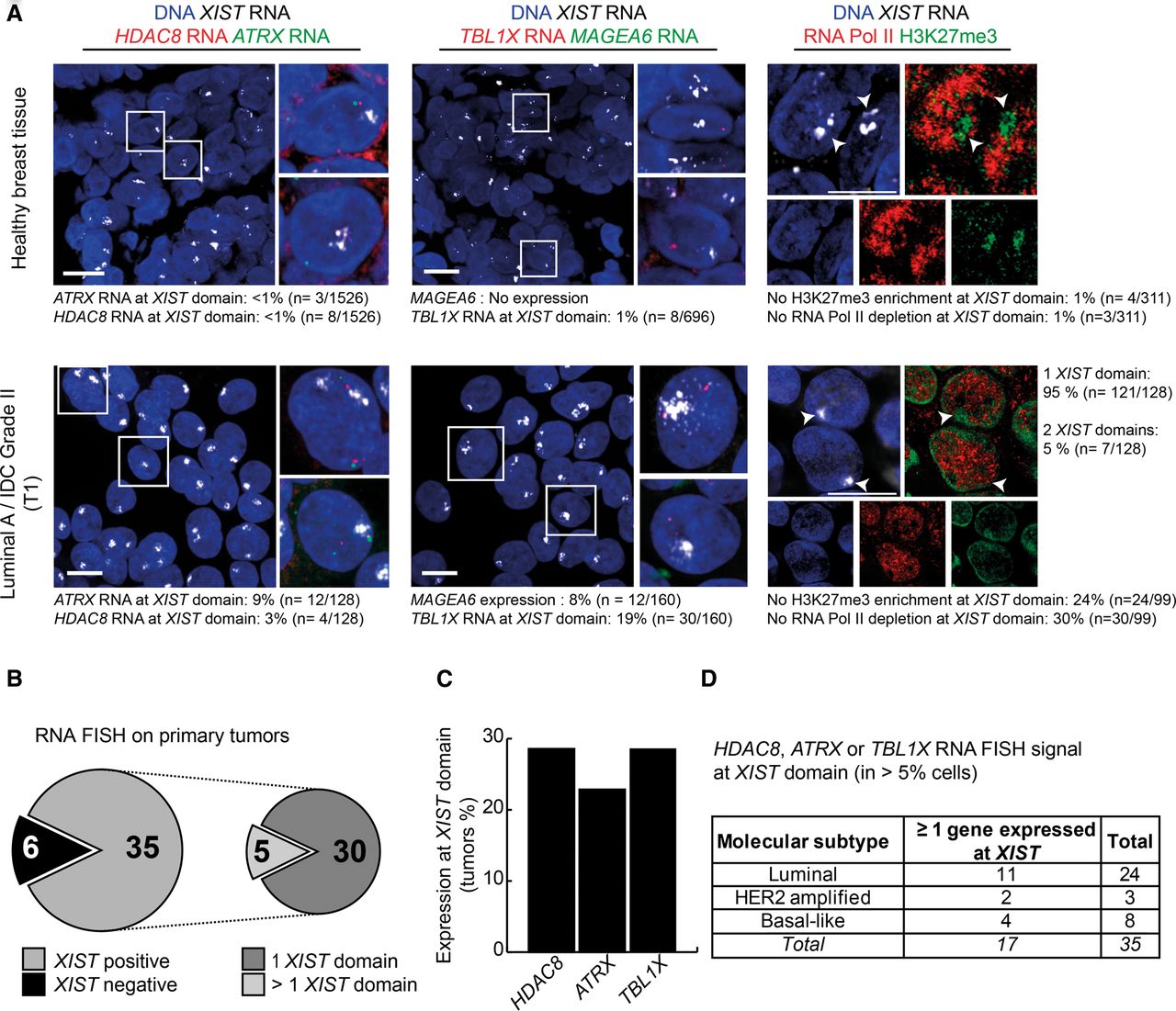
The inactive X chromosome is reactivated in primary breast tumors. (A) Z-projections of 3D RNA FISH show representative examples of expression of HDAC8 (red) and ATRX (green) (left) or TBL1X (red) and MAGEA6 (green) (middle) at XIST domains (gray) in healthy breast tissue and invasive ductal carcinoma (IDC; Luminal A Grade III tumor). On the right, Z-projections of super-resolutive 3D immuno-RNA FISH show representative examples of the level of H3K27me3 enrichment (green) and RNA Pol II depletion (red) on XIST RNA domains (gray) in healthy and tumoral breast tissues. Arrowheads indicate the XIST domains. Quantification of RNA Pol II exclusion and H3K27me3 enrichment at XIST domains have been carried out on images acquired with a confocal spinning-disk microscope. Scale bar, 10 µm. (B) Summary of the XIST domain positive (domains in >10% of the nuclei) and negative tumors among the 41 primary breast tumors studied. (C) Summary of the number of tumors harboring HDAC8, ATRX, or TBL1X expression at XIST domain (assessed by RNA FISH). A gene showing expression within the XIST domain in >5% of the nuclei is considered as reactivated in this tumor. (D) The table recapitulates the number of XIST positive tumors with Xi-linked gene reactivation according to their molecular subtypes: Luminal, HER2 amplified, or Basal-like (BCL).


