
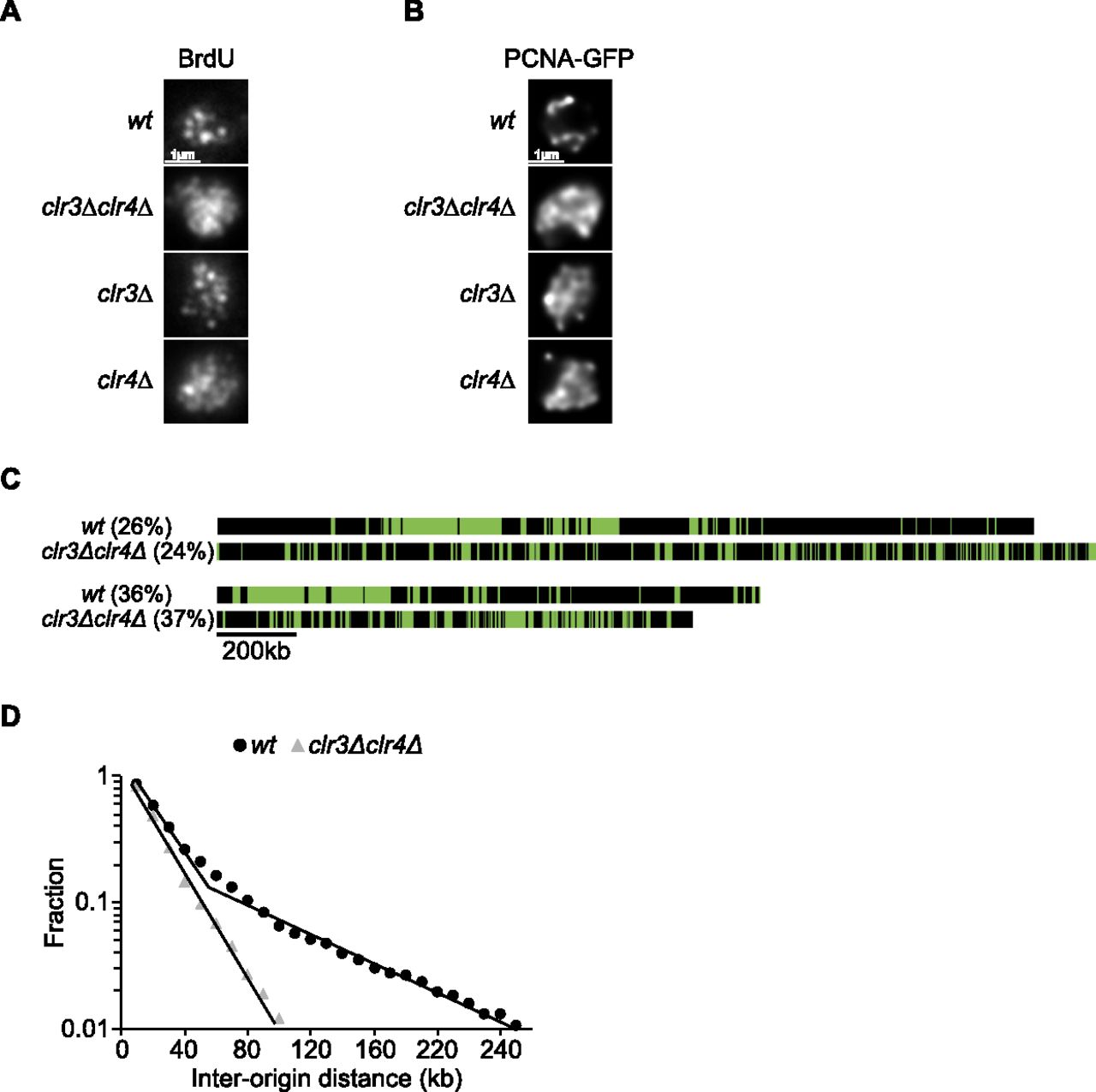
A clr3∆clr4∆ mutant disrupts both the organization of replication foci within the nucleus and clusters with fired origins along the chromosomes. (A) Immunostaining of BrdU-labeled replication foci in wt, clr3∆, clr4∆, and clr3∆clr4∆ cells. Images show an equatorial optical section. (B) Live imaging of PCNA-GFP-labeled replication foci. Images show an equatorial optical section. (C) Comparison between wt and clr3∆clr4∆ molecules at the same percentage of DNA replication. Clusters are present in wt molecules but are not observed in clr3∆clr4∆ molecules. (D) Semilog plot of the cumulative frequencies of IODs for molecules replicated from 20% to 50% for wt and clr3∆clr4∆ cells. The values on the y-axis correspond to the fraction of IODs that are larger in size than the corresponding IOD on the x-axis. The data points for clr3∆clr4∆ cells are fitted with a single straight line. The second straight line with the shallow slope described for wt cells is not present in clr3∆clr4∆ cells.


