
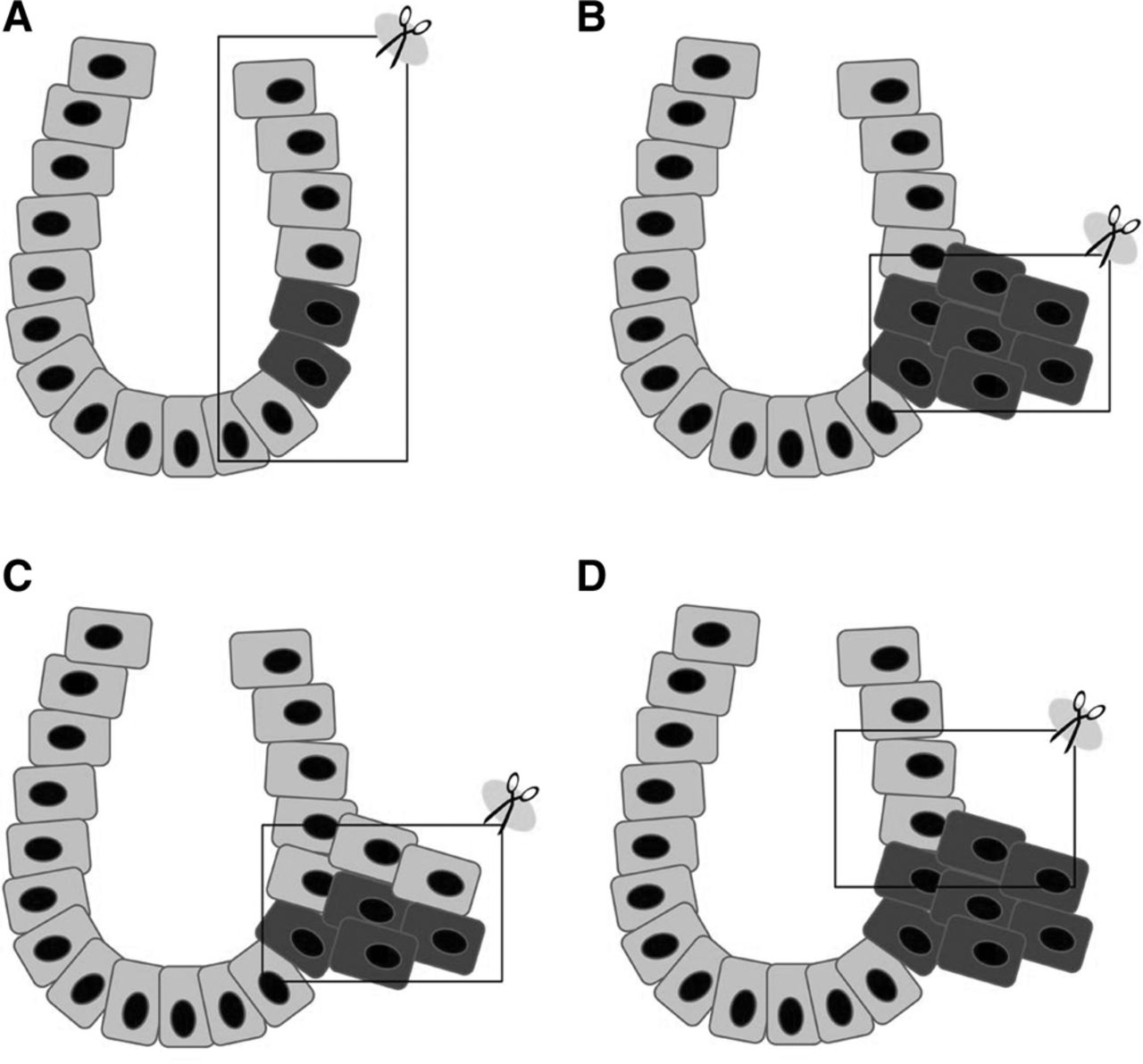
Figure 4.
Distribution of somatic L1 insertions. (A) Normal colon crypt containing a few cells with L1 insertions (detectable only by nested PCR). (B) Colon tumor with early L1 insertions (detectable by conventional PCR). (C) L1 insertions occurring late during tumorigenesis (detectable only by nested PCR). (D) Colon tumor containing early L1 insertions with contaminating normal cells (detectable only by nested PCR). Note that by using nested PCR, it may be possible to misdiagnose contaminating or tumor-invading normal tissue-specific insertions (A) as tumor-specific (C or D). The frame represents the sampled tissue.


