
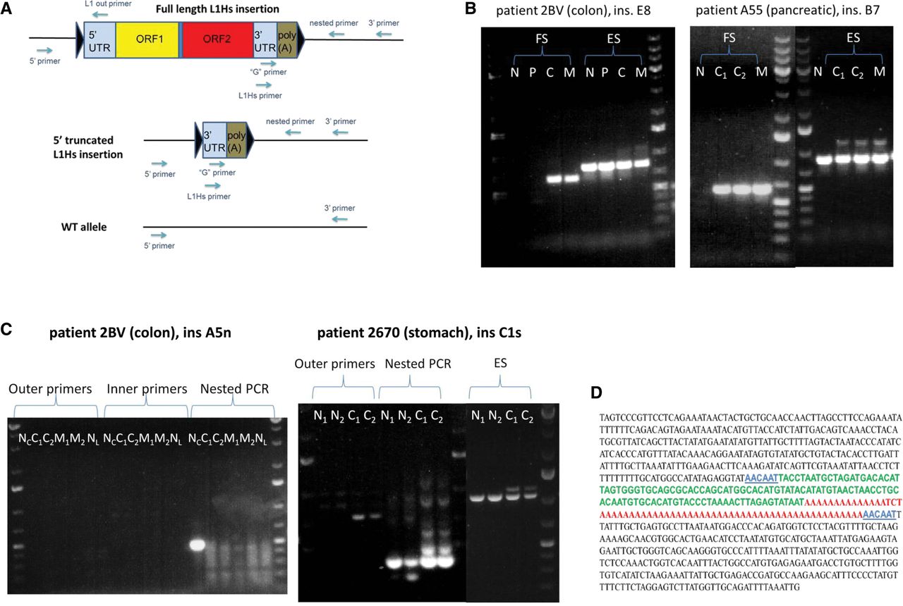
PCR and Sanger-sequencing validation scheme of L1-seq results. (A) Multistep PCR validation scheme and location of primers used. Insertions were primarily validated with conventional PCR at their 3′ junction using the L1Hs with the 3′ primer. Some insertions were also validated with nested PCR using the “G” primer with a nested 3′ primer. After the 3′ junction was located, we attempted to find the 5′ junction using the 5′ primer with L1 out primers. Triangles symbolize target site duplications (TSD). (B) PCR validation of clonal cancer-specific insertions. (Left panel) A primary colon cancer-and-metastasis-specific insertion (ins. E8). (Right panel) A primary pancreatic cancer-and-metastasis-specific insertion (ins. B7). The higher molecular weight bands visible above the tumor tissues of the empty site PCR products are the highly truncated L1 elements, as assessed by gel extraction and Sanger sequencing. (N) Normal, (P) polyp, (C) primary cancer, (C1) primary cancer section 1, (C2) primary cancer section 2, (M) metastasis, (FS) filled site PCR product (insertion allele), (ES) empty site PCR product (wild-type allele). (C) PCR validation of the normal colon-specific insertion “A5n” and the somatic normal-and-cancer-specific insertion “C1s” in stomach cancer. “A5n” is detectable exclusively using nested PCR in case 2BV, while the somatic L1 insertion in the stomach cancer of patient 2670 is detectable by both conventional and nested PCR and is also detectable in normal stomach using nested PCR. (NC) Normal colon, (NL) normal liver, (N1) normal stomach section 1, (N2) normal stomach section 2, (C1) primary cancer section 1, (C2) primary cancer section 2, (M1) metastasis section 1, (M2) metastasis section 2. O'GeneRuler 1 kb Plus DNA ladder was used (Thermo Scientific). (D) Reconstituted Sanger sequence of the 5′ truncated colorectal cancer-and-metastasis-specific ins. E8 from B. In blue, TSD (6 bp, alternatively, 7 bp due to microhomology at the 5′ junction); in green, highly truncated L1Hs (112/112-bp identity with L1RP, nt 5908–6019); in red, poly(A) tail.


