
Figure 1.
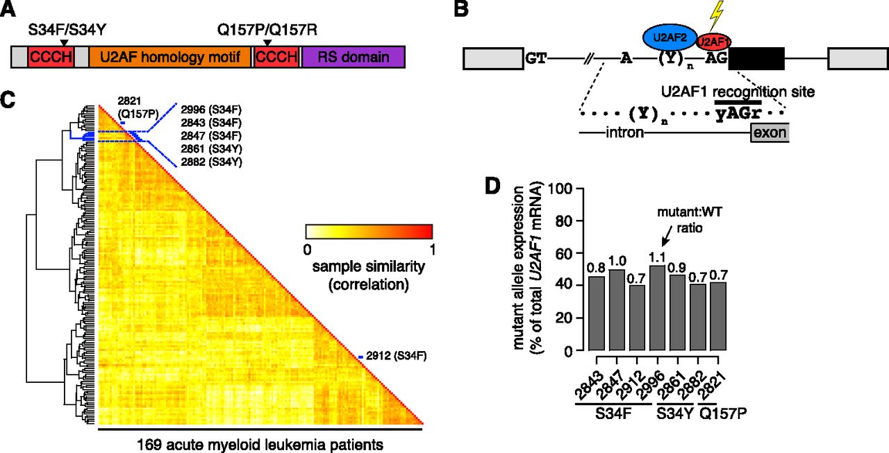
U2AF1 mutations contribute to splicing programs in AML. (A) U2AF1 domain structure (Kielkopf et al. 2001; The UniProt Consortium 2012) and common mutations. (CCCH) CCCH zinc finger. (B) Schematic of U2AF1 interaction with the 3′ splice site of a cassette exon (black). (C) Heat map illustrating similarity of alternative splicing programs in AML transcriptomes. Dendrogram is from an unsupervised cluster analysis based on cassette exon inclusion levels. (Blue) Samples with U2AF1 mutations. (D) U2AF1 mutant allele expression as a percentage of total U2AF1 mRNA in AML transcriptomes. Numbers above bars indicate the ratio of mutant to WT allele expression.


