
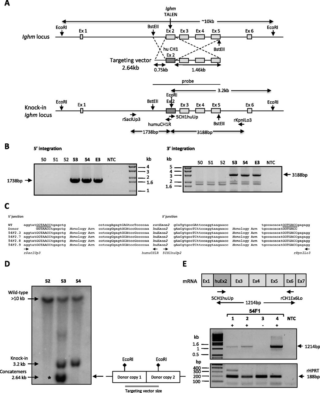
Targeted exon exchange into the rat Ighm locus. (A) (Upper) Diagrams showing schematic representations of the rat Ighm locus with the site of TALE nuclease action and of the targeting vector containing exon 2 of human IGHM flanked by 0.75-kb-long 5′ and 1.46-kb-long 3′ homology arms. (Lower) Diagram showing the integration by homologous recombination of the donor DNA sequence in the targeted locus, and the position of primers used for 5′ and 3′ junction PCRs. Integration in the 5′ and 3′ sides should generate fragments of 1738 bp and 3188 bp, respectively. EcoRI is the restriction enzyme used in Southern blot analyses due to the presence of the EcoRI site in the human but not in the rat Ighm sequence. The probe used in Southern blot analyses is the targeting vector. In the case of homologous recombination events, a 3.2-kb band is expected. (B) The left and right gels show the results analyzing, respectively, the 5′ and 3′ extremity of DNA integration, using the pair of primers indicated in A. Three animals (53, 54, and E3) showed a band of expected size (1738 bp in 5′ and 3188 bp in 3′ ends). (NTC) No template control. (C) Sequence comparison at the 5′ and 3′ junctions with wild-type genomic DNA and donor DNA sequences of four representative F2s (54F2.3, 54F2.7, 54F2. 8, and 54F2.9: F2 generation from animal 54). Exon exchange was confirmed at the BstEII site in 5′ and 3′ (underlined). (D) Southern blot analysis of founders for homologous recombination integration. Genomic DNA was digested with EcoRI and 10 µg of DNA were loaded per lane. Blots were probed with the targeting sequence, as indicated in A. Arrows indicate bands of 10 kb and 3.2 kb corresponding to wild-type sequences and HDR sequences, respectively. An asterisk marks the presence of concatemers showing a band at 2.64 kb (the size of the transgene). The diagram at the right explains the expected size of the concatemers once linearized. Two animals (founders 53 and 54) harbor a HDR insertion, whereas no HDR event is observed in the third (animal 52). More than one copy of the donor DNA sequence is observed in animal 53. (E) (Upper) Diagrams showing a schematic representation of the mRNA sequence in HDR animals and the position of primers used for RT-PCR. A band of 1214 bp is expected. (Lower) Electrophoresis gel pictures show the presence of an amplified band of 1214 bp in three animals born from the mating of the founder 54 (HDR) with a wild-type rat. The amplification of rat HPRT serves as a control. (NTC) No template control.


