
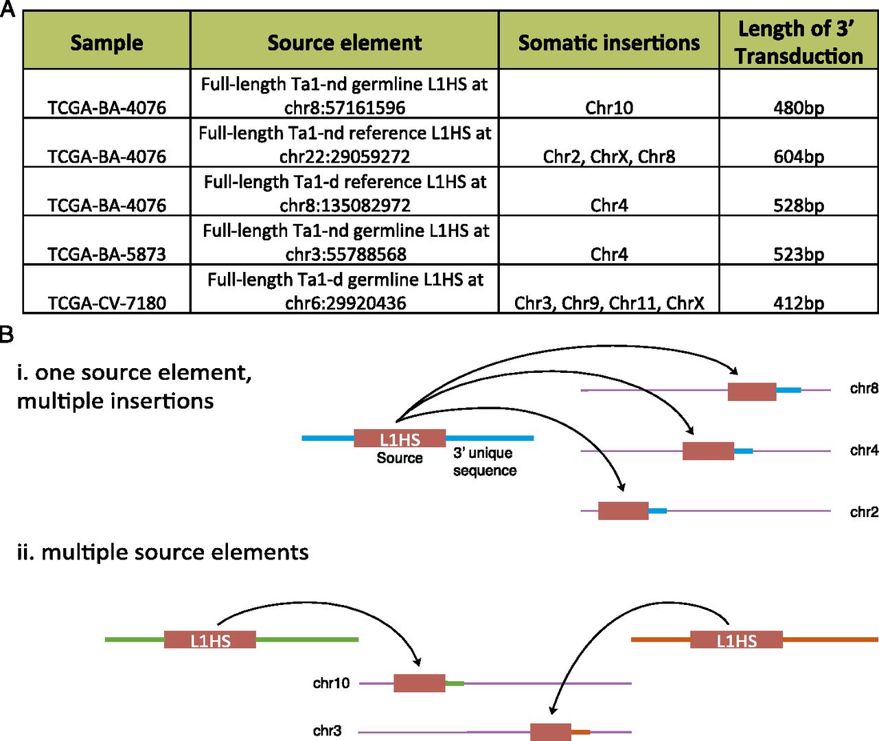
Figure 3.
3′-transductions elucidate source retrotransposon element. (A) Select 3′-transduction events, including the sample, the source element location (i.e., genomic origin of the unique sequence), the transposition insertion location, and the length of the transduced sequence. See Supplemental Table 4 for a full list. (B) Schematic of the two models of somatic retrotransposition detected seen in this analysis: (i) one source L1HS element becoming active and inserting multiple times across the tumor sample, and (ii) several source elements becoming active in the tumor sample.


