
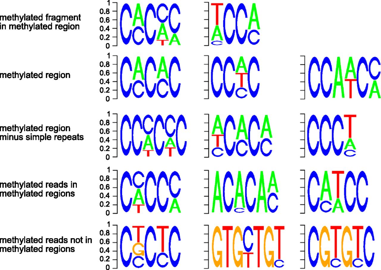
Sequence motifs in which methylation occurs. The motifs were identified by DREME analysis of different types of methylated sequences. The row at the top was derived using sequences identified within methylated regions as containing a high density of methylated cytosine; the first motif is found in 57% of these methylated fragments, and the second motif in 33% of fragments (Table 2). The second row was derived using the entire sequence of each of the 25,319 methylated regions. These motifs are highly similar to the motifs shown in the top row, which were derived from fragments (short sequences) within methylated regions. The third row was derived using the same entire methylated regions as in the top row, but excluding all regions overlapping with a simple sequence repeat. The similarity between the top and second rows indicates that the motifs associated with methylation are not confined to simple sequence repeats. The fourth row was derived using only sequence reads that contain at least one methylated cytosine, and align to a methylated region. Again, these motifs are similar to the others above, further corroborating the association of the motif with methylation; all these motifs are devoid of guanine. The enrichment of methylated regions in simple sequence repeats is largely explained by the presence of these motifs in some simple sequence repeats (Supplemental Fig. S10). Finally, the bottom row was derived using sequence reads that do not align to a methylated region but contain at least one methylated cytosine. The contrast between these motifs and those derived from methylated reads within methylated regions (fourth row) supports the specificity of the motif associated with methylated regions. It may also suggest the presence of a much rarer type of methylation associated with a different sequence motif.


