
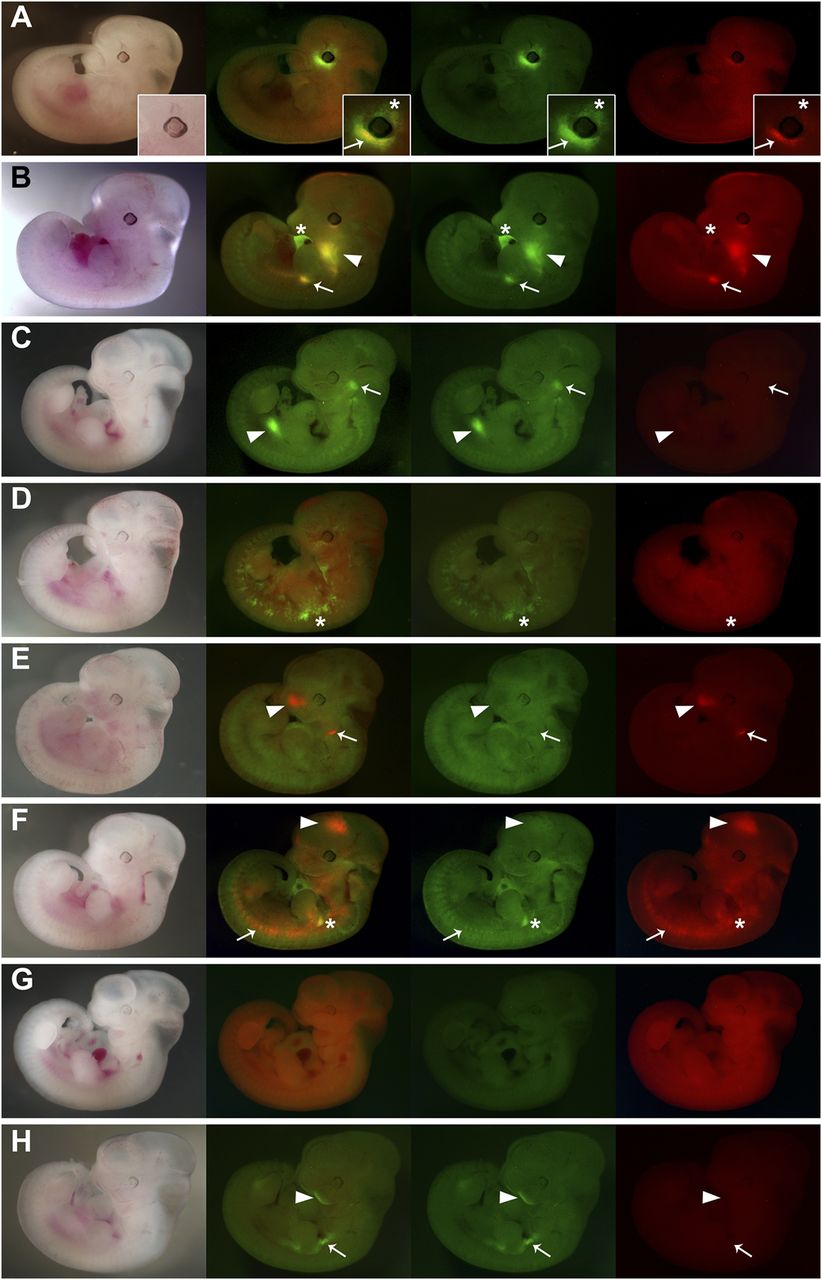
Mouse transgenic ED lines. Eight mouse transgenic embryos were generated using the ED system. (A) In this example, GFP expression is detected in the dorsal periocular mesenchyme (asterisk; inset), and coexpression of GFP and RFP is observed in the ventral periocular mesenchyme (arrow; inset). (B) Strong GFP expression is detected in the heart (asterisk), while GFP and RFP coexpression is detected at the base of the forelimb (arrow), the branchial arches (arrowhead), and at low levels in the somites. (C) GFP expression is detected in the trigeminal ganglion (arrow) and the midgut (arrowhead), while RFP expression is not detected. (D) This embryo expresses GFP throughout the embryo in migratory cells that probably correspond to dermis (asterisk), while RFP is found ubiquitously at low levels. (E) RFP expression is detected in the olfactory pits (arrowhead) and the second branchial arch (arrow), possibly in the mesodermal core, while GFP is detected at low levels ubiquitously. (F) RFP expression alone is detected in the forebrain (arrowhead), somites (arrow), and cells migrating into the limb buds; RFP and GFP coexpression is strongly detected in the proximal region of the forelimb (asterisk), while unique GFP expression is detected at low levels ubiquitously. (G) GFP and RFP are coexpressed ubiquitously at low levels. (H) Expression of GFP alone is observed superficially in the mandibular component of the first branchial arch (arrowhead), in tissues surrounding the fore and hindlimbs (arrow), and RFP expression is not detected. The stages of the embryos shown are 11.5 d post-coitum (dpc; A,B) and 10.5 dpc (C–H). The first column presents transmitted light images, the second an overlay of GFP and RFP channels, the third the GFP channel alone (green), and the fourth the RFP channel alone (red).


