
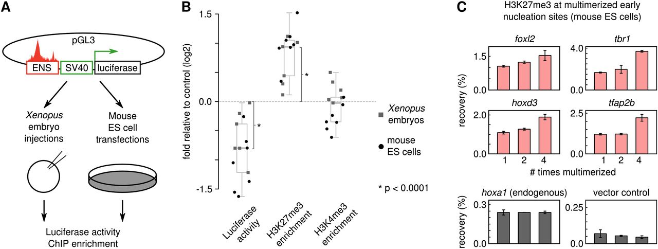
Early H3K27me3 nucleation sites encode conserved repressive capacity. (A) Repression assay of H3K27me3 early nucleation sites in Xenopus embryos and mouse ES cells. (B) Early nucleation sites function as repressive elements. Shown is luciferase activity normalized to Renilla as fold change relative to control, H3K27me3 ChIP-qPCR enrichment relative to control, and H3K4me3 ChIP-qPCR enrichment relative to control (log2 scale; significance, Wilcoxon signed rank test; Xenopus, light square dots; ES cells, dark round dots). (C) H3K27me3 enrichment increases with length of susceptible sequence. Shown is the H3K27me3 ChIP-qPCR recovery for four SVM-positive H3K27me3 nucleation sites, an endogenous locus (hoxa1), and a vector backbone control.


